Abstract
Objective
The aim of the present study was to evaluate the effects of PRP on Achilles tendon healing in rabbits during the inflammatory, proliferative, and remodeling phases by histological examination and quantitative assessments.
Methods
Fifty mature male Japanese albino rabbits with severed Achilles tendons were divided into two equal groups and treated with platelet-rich plasma (PRP) or left untreated. Tendon tissue was harvested at 1, 2, 3, 4, and 6 weeks after treatment, and sections were stained with hematoxylin-eosin and monoclonal antibodies against CD31 and type I collagen.
Results
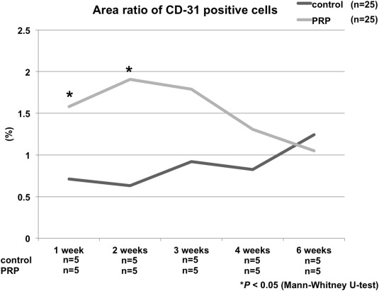
Collagen fibers proliferated more densely early after severance, and subsequent remodeling of the collagen fibers and approximation of normal tendinous tissue occurred earlier in the PRP group than in the control group. The fibroblast number was significantly higher in the PRP group than in the control group at 1 and 2 weeks. Similarly, the area ratio of CD31-positive cells was significantly higher in the PRP group than in the control group at 1 and 2 weeks. Positive staining for type I collagen was more intense in the PRP group than in the control group after 3 weeks, indicating tendon maturation.
Conclusion
Administration of PRP shortened the inflammatory phase and promoted tendon healing during the proliferative phase.
Keywords
Achilles tendon ; Growth factor ; Platelet-rich plasma ; Rabbit ; Tendon healing
Introduction
Current treatments for acute Achilles tendon rupture with or without surgery involve early accelerated rehabilitation, which shortens the immobilization period and permits early range of motion and muscle strengthening exercises.1 ; 2 Using this approach, multiple studies have reported immediate weight-bearing capability after injury.3 ; 4 Experimental models suggest that early mobilization and loading of the severed Achilles tendon promotes histological maturation of collagen fibers.5 ; 6 ; 7 ; 8 It is likely that, in the future, Achilles tendon rupture will be treated not only by mechanical stimuli such as early weight-bearing and range-of-motion exercises, but also by biological approaches such as growth factor treatment.9
Tendon healing comprises mutually dependent and overlapping inflammatory, proliferative, and remodeling phases. In the initial inflammatory phase, erythrocytes and inflammatory cells enter the injury site, and necrotic tissue is phagocytosed by monocytes and macrophages. Growth factors are released, which in turn stimulate angiogenesis and proliferation of fibroblasts, which migrate to the injury site and initiate type III collagen synthesis. The proliferative phase begins a few days later; at this time, type III collagen synthesis is maximal and remains elevated for approximately 6 weeks. During the subsequent remodeling phase, a higher proportion of type I collagen is synthesized, and tenocytes and collagen fibers become aligned in the direction of stress.10 ; 11
Platelet-rich plasma (PRP) comprises autologous blood with platelet concentrations above baseline values. PRP can potentially promote the healing process by delivering various growth factors contained in α-granules, which play important roles in cell proliferation, chemotaxis, cell differentiation, and angiogenesis.12 ; 13
To the best of our knowledge, the effects of PRP on the three phases (inflammatory, proliferative, and remodeling phase) of tendon healing have not been investigated. The aim of the present study was to evaluate the effects of PRP on Achilles tendon healing in rabbits during the three phases by histological examination and quantitative assessments. We hypothesized that PRP treatment promotes tissue healing by stimulating fibroblast proliferation and collagen production, thereby accelerating the remodeling process.
Materials and methods
All experiments were approved by the Institutional Animal Care and Use Committee of our institution. Male Japanese albino rabbits (n = 50) weighing 3 kg were housed at 22–24 °C on a 12:12-h light/dark cycle with free access to water and a standard diet.
Preparation of PRP
Four male Japanese white rabbits were used for PRP production. PRP was prepared using the commercial Cascade® Autologous Platelet System (MTF Sports Medicine/Cascade Medical Enterprises, Wayne, NJ, USA) according to the manufacturers recommendations.14
Achilles tendon rupture model
The Achilles tendon rupture model was established as described previously.8 Briefly, rabbits were anesthetized with sodium pentobarbital (20 mg/kg body weight intravenously), and the skin of right hind limb shaved. The surgical procedure was performed under the aseptic condition. A longitudinal skin incision was made in the midline of the Achilles tendon. The paratenon was cut in the same direction as that of the skin incision, and the Achilles and plantaris tendons were dissected from the surrounding tissues. The total length of Achilles tendon was about 3 cm. The middle of the Achilles tendon was severed using a scalpel (approximately 1.5 cm proximal to the calcaneus insertions). The plantaris tendon was left intact as an internal splint that maintained a very short distance (0–1 mm) between the two stumps of the Achilles tendon. The severed tendon was not sutured.
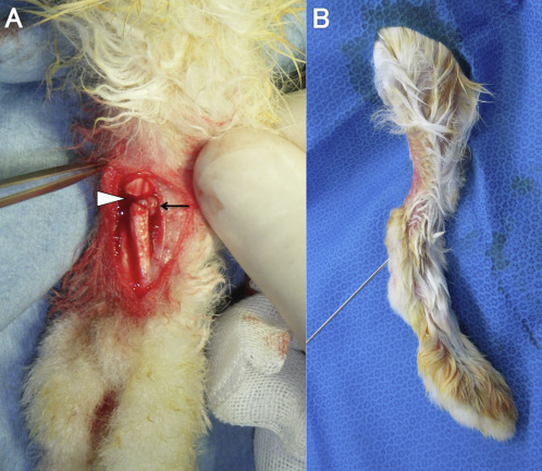
The rabbits were divided into two groups (n = 25 each). The control group underwent only severance of the Achilles tendon; in the PRP group, 0.3 g PRP was placed into the anterior side of the tendon stumps, immediately after cutting the tendon (Fig. 1 A); then, the paratenon and skin were sutured. After the surgery, the ankle was immobilized at 60° plantar flexion in both groups by percutaneous insertion of a 1.8-mm Kirschner wire from the calcaneus through the talocrural articulation to the tibia (Fig. 1 B); immobilization was supplemented with a short leg cast. All rabbits were observed in conventional cages made of wire cloth. Postoperative immobilization was continued until the Achilles tendon harvesting was completed.
|
|
|
Fig. 1. Surgical procedure. (A) Stumps of the Achilles tendon (white arrow head) and platelet-rich plasma: PRP (black arrow). (B) Ankle was immobilized at 60° plantar flexion by percutaneous insertion of a Kirschner wire. |
Histological examination
Achilles tendons were harvested 1, 2, 3, 4, and 6 weeks after surgery by dissection. The number of specimens was 5 at each week, in both the control and PRP groups. The tissue was fixed in 20% buffered neutral formalin for 3 days, dehydrated, and then embedded in paraffin. Longitudinal sections of the tendon were stained with hematoxylin and eosin (H&E). For immunohistochemistry, the Achilles tendon was sectioned in the longitudinal plane at a thickness of 7 μm. Sections were incubated with monoclonal antibodies against cluster of differentiation (CD) 31 (Dako Corp., Carpinteria, CA, USA)—which labels endothelial cells—and type I collagen (Abcam, Cambridge, UK). Immunohistochemical staining was carried out using an Envision™+ kit (Dako Corp.). Sections were counterstained with Mayers hematoxylin.
The tendon healing process was compared between the two groups by evaluating the results of H&E staining. Type I collagen immunoreactivity was evaluated semi-quantitatively using the scoring system shown in Table 1 .15 The H&E staining results and CD31 immunoreactivity were quantified by analyzing images of the gap between the Achilles tendon stumps that were acquired with a BX50 light microscope (Olympus, Tokyo, Japan) equipped with a DP72 digital camera (Olympus). Proliferation was evaluated by counting the number of fibroblasts in 10 fields at 400× magnification. To assess angiogenesis, we measured the area ratio of CD31-positive cells in 10 fields at 100× the original magnification using the Lumina Vision v.2 image analysis software (Mitani Corp., Tokyo, Japan).
| Criterion for staining | Score |
|---|---|
| No staining | 0 |
| Faint | 1 |
| Patchy | 2 |
| Medium | 3 |
| Medium-to-strong | 4 |
| Strong | 5 |
Results are expressed as mean ± standard deviation. Significant differences between groups were evaluated with the Mann–Whitney U-test using SPSS v.12.0 (SPSS Inc., Chicago, IL, USA). P < 0.05 was considered statistically significant.
Results
The mean platelet concentration in whole blood was 40.6 ± 9.6 × 10⁴ (mean ± standard deviation)/μL. The mean platelet concentration in the PRP was 105.7 ± 22.8 × 10⁴ /μL, which was approximately 2.6-times greater than that in whole blood.
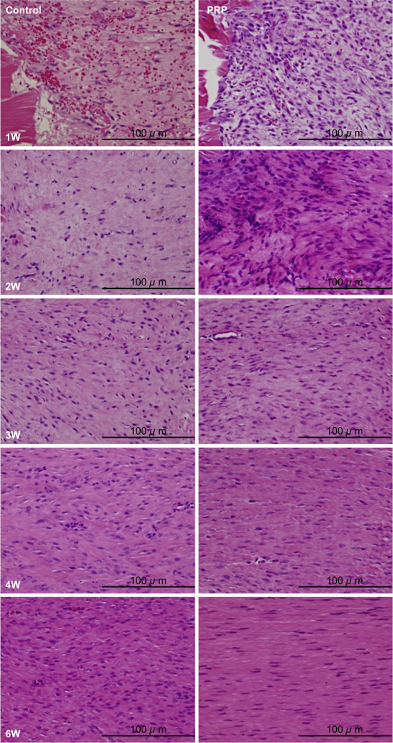
One week after surgery, the gap between the stumps of the severed Achilles tendon was found to be filled with fibroblasts, blood cells, and fibrin in both PRP-treated and untreated rabbits; however, no increase in collagen fiber density was observed. Collagen fibers were sparse at 2 weeks in the control group; however, in the PRP group, a dense network of fibers and capillaries was present in the gap. At 3 weeks, collagen fiber density was increased in both groups. At 4 weeks, fiber bundles were not oriented along the axis of the tendon, and fibroblasts continued to proliferate in the control rabbits. In contrast, in the PRP-treated rabbits, collagen fiber bundles were aligned in a single direction parallel to the long axis of the tendon. At 6 weeks, collagen fiber bundles were oriented along the axis of the tendon partially in the control rabbits. In contrast, in the PRP-treated rabbits, collagen fiber bundles were aligned parallel along the axis of the tendon, and they closely approximated fibers in normal tendons (Fig. 2 ).
|
|
|
Fig. 2. Hematoxylin–Eosin staining (original magnification 400×). Greater density of collagen fibers was observed at earlier time points, and fibers were aligned in a more orderly fashion at later time points in PRP-treated rabbits compared with control rabbits. |
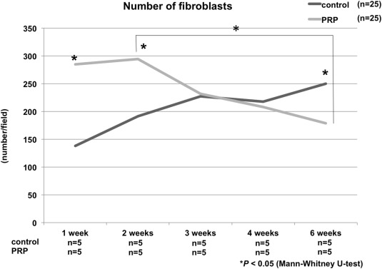
Fibroblast migration increased gradually in both groups starting from week 1, but more fibroblasts were observed in the PRP group than in the control group at 1 and 2 weeks (P = 0.010 and P = 0.009, respectively). The number of fibroblasts remained high in the control group at 3 weeks; however, in the PRP-treated rabbits, there were more mature cells with flattened nuclei than proliferating fibroblasts with spindle-shaped nuclei at 3 and 4 weeks (P = 0.853 and P = 0.481, respectively; Fig. 3 ), indicating a decrease in the number of immature fibroblasts. At 4–6 weeks, fibroblast proliferation persisted in the control group; however, during this period, the number of fibroblasts decreased markedly (P = 0.001), and more fibroblasts with flattened nuclei were observed in the PRP group ( Fig. 2 ). In the PRP-treated rabbits, the number of fibroblasts decreased significantly at 6 weeks compared to that at 2 weeks (P = 0.001), whereas that in the control groups continued to increase at 6 weeks ( Fig. 3 ).
|
|
|
Fig. 3. The number of fibroblast was higher at 1 and 2 weeks but lower at 6 weeks in rabbits treated with PRP than in controls. *P < 0.05. |
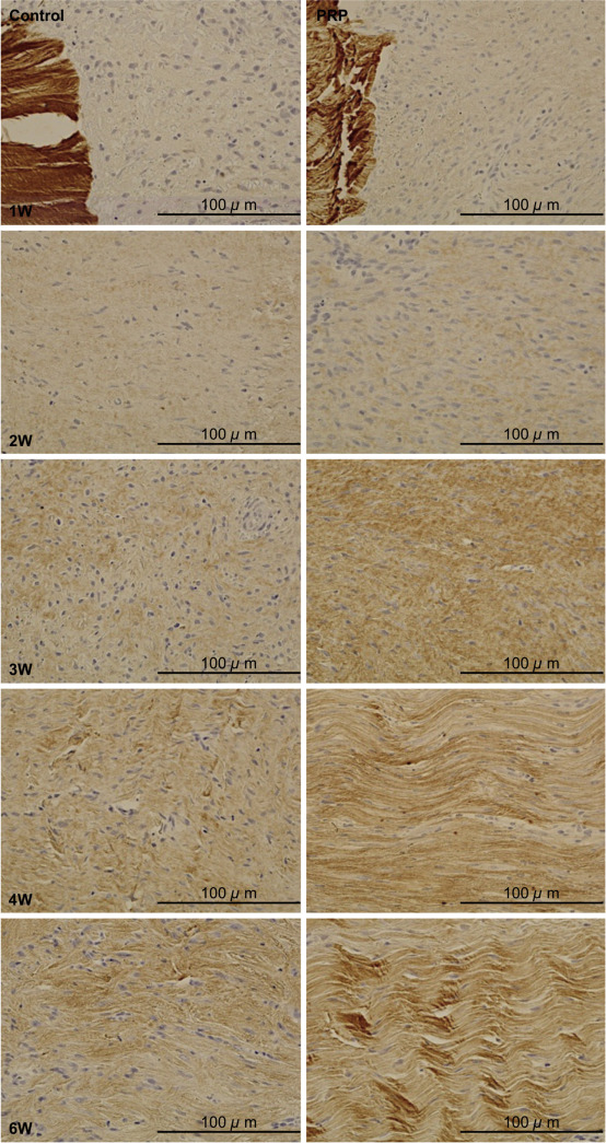
No type I collagen immunoreactivity was observed in either group until week 2 (staining intensity score = 1). At 3 weeks, the signal intensity was higher in the PRP-treated group than in the control group (score = 3 vs. 2), indicating that the tendons were more mature in the former at this time point. Accordingly, type I collagen immunoreactivity scores at 4 and 6 weeks were 2 and 3, respectively, for the control group, but were 3 and 4, respectively, for the PRP-treated rabbits (Fig. 4 ).
|
|
|
Fig. 4. Type I collagen expression (original magnification 400×). Type I collagen immunoreactivity was started from 3 weeks, and signal intensity was greater in PRP-treated rabbits than in control rabbits. |
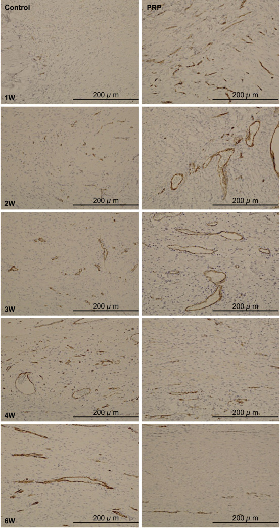
Vascular proliferation, as evidenced by increased blood vessel diameter, was higher in the PRP group than in the control group at 1 and 2 weeks. Vessel diameter increased in the control group at 3 and 4 weeks, as did the number of the blood vessels; in contrast, at this time point, vessel diameter and the number of vessels decreased in the PRP group. At 6 weeks, blood vessels had smaller diameters and were scattered throughout the healing tendon in the control group; however, in the PRP group, blood vessels were only observed between collagen fiber bundles (Fig. 6 ).
|
|
|
Fig. 5. CD31 expression (original magnification 200×). A greater number of cells and larger vessel diameters were observed in PRP-treated rabbits than in control rabbits at 1 and 2 weeks. |
|
|
|
Fig. 6. The area ratio of CD31-positive cells was higher in PRP-treated rabbits than in control rabbits at 1 and 2 weeks; in the PRP group, CD31-positive cells decreased after reaching a peak at 2 weeks. *P < 0.05. |
The area ratio of CD31-positive cells was significantly higher in the PRP group than in the control group at 1 and 2 weeks (P = 0.023 and P = 0.011, respectively). At 3, 4, and 6 weeks, the area ratios between the two groups were similar (P = 0.052, 0.190, and 0.529, respectively); however, the value reached a peak at 2 weeks in the PRP group but remained high in the control group until 6 weeks ( Fig. 5 ).
Discussion
The number of fibroblasts and degree of neovascularization were higher in the PRP-treated group than in the control group at 1 and 2 weeks after injury. This result suggests that PRP promotes fibroblast migration and/or proliferation as well as neovascularization in the early stages of tendon healing. The number of fibroblasts and area ratio of CD31-positive cells decreased after reaching a peak at 2 weeks in the PRP-treated rabbits, suggesting initiation of the remodeling phase. Fibroblast numbers were markedly reduced in the PRP group at 6 weeks; consistent with these changes, collagen fiber bundles were aligned in one direction, parallel to the long axis of the tendon. These results suggest that PRP treatment hastened the initiation of the remodeling phase and promoted tendon tissue repair, including more rapid maturation of scar tissue between tendon stumps and return of tendon tissue to the normal state.
On the other hand, in untreated rabbits, neovascularization and fibroblast proliferation persisted 6 weeks after injury, suggesting that, in the absence of exogenous growth factors, fibroblasts remained in a proliferative state, although some remodeling may have been initiated at this time. Type I collagen expression was also lower in the untreated rabbits than in those treated with PRP, indicating slower tendon maturation. These findings are consistent with those of a previous study reporting that the remodeling phase was initiated and cellularity was decreased approximately 6 weeks after Achilles tendon injury.11
We propose the following mechanism to explain the effects PRP on tendon healing. In the inflammatory phase, growth factors released from PRP stimulate fibroblast migration and proliferation as well as neovascularization. The large number of migrating fibroblasts leads to early initiation of the proliferative phase and increased production of collagen fibers. Consequently, the remodeling phase is initiated earlier than in untreated rabbits. Tension aligns fibroblasts parallel to the line of force.5 ; 8 The application of tension to healing tissue by knee joint mobilization and immediate weight-bearing in the remodeling phase may have caused parallel alignment of collagen fiber bundles in the early stages of healing in the PRP-treated rabbits,8 yielding regenerated tissue that more closely approximated normal, uninjured tendon tissue.
In animal models, many studies have provided evidence for the beneficial effects of PRP on tendon healing.16 ; 17 On the other hand, some studies concluded that PRP treatment is not useful for Achilles tendon rapture.18 ; 19 In this way, the effect of PRP is still unclearly also in animal experiments. PRP has been used in many clinical applications, including in the treatment of acute rupture of the Achilles tendon, however, the effect of this treatment for Achilles tendon rupture is controversial. Sánchez et al have suggested that operative management of Achilles tendon tears associated with the application of autologous platelet-rich fibrin could present new possibilities for enhanced healing and functional recovery.20 In contrast, Schepull et al have suggested that there is no dramatic positive effect of PRP in healing of Achilles tendon rupture and that it has a negative effect on the functional 1-year result.21 However, the postoperative treatment in those two studies was different. Sánchez et al used a below-knee cast for 2–3 weeks, and patients were allowed to walk for this period. On the other hand, Schepull et al used a below-knee cast after 7 weeks, and full weight bearing was allowed only after 3.5 weeks. These differences in postoperative treatment may have affected the functional outcome. We believe that PRP treatment combined with accelerated rehabilitation is essential for the treatment of acute Achilles tendon rupture.
It is clinically recommended that Achilles tendon rupture be treated with early weight bearing and a shortened immobilization period to re-establish a range of motion as early as possible.22 ; 23 We propose that shortening the inflammatory and proliferation phases and accelerating the initiation of the remodeling phase is crucial for the treatment of acute Achilles tendon rupture. The inflammatory and proliferation phases often correspond with the initiation of early-accelerated rehabilitation in the clinical treatment of acute Achilles tendon rupture. Moreover, because the strength of the tendon is low in these phases, accelerated rehabilitation during this period could potentially lead to elongation or re-rupture of the healing tendon. Accordingly, PRP treatment promotes tendon healing to facilitate safe, accelerated rehabilitation throughout this period.
There were a few limitations to the present study. First, we used the commercial Cascade® Autologous Platelet System to prepare PRP, which may have delayed the release of growth factors by creating a gel-like PRFM and thereby complicated the direct measurement of platelet concentration. Therefore, we determined the platelet concentration in the PRFM from the difference between the number of platelets in the liquid PRP produced in the first centrifugation step and that in the platelet-poor plasma (PPP) produced in the second centrifugation step. Second, we didn't conduct mechanical tests of healing tendon. In this study, we can't determine mechanical properties of healing tendon and biomechanical effect of PRP treatment.
In conclusion, administration of PRP shortened the inflammatory phase and promoted tendon healing during the proliferative phase. The remodeling phase began earlier with PRP treatment, and earlier approximation of normal tendon tissue in the healing tissue was facilitated. Our findings indicate that PRP treatment combined with early functional rehabilitation can accelerate Achilles tendon healing without tendon elongation or re-rupture and thereby reduce the requisite downtime from work or sports activities.
References
- 1 B.C. Twaddle, P. Poon; Early motion for Achilles tendon ruptures: is surgery important? A randomized, prospective study; Am J Sports Med, 35 (2007), pp. 2033–2038
- 2 D.M. van der Eng, T. Schepers, N.W.L. Schep, J.C. Goslings; Rerupture rate after early weightbearing in operative versus conservative treatment of Achilles tendon ruptures: a meta-analysis; J Foot Ankle Surg, 52 (2013), pp. 622–628
- 3 S.W. Young, A. Patel, M. Zhu, et al.; Weight-bearing in the nonoperative treatment of acute Achilles tendon ruptures: a randomized controlled trial; J Bone Joint Surg Am, 96 (2014), pp. 1073–1079
- 4 K.W. Barfod, J. Bencke, H.B. Lauridsen, I. Ban, L. Ebskov, A. Troelsen; Nonoperative dynamic treatment of acute Achilles tendon rupture: the influence of early weight-bearing on clinical outcome: a blinded, randomized controlled trial; J Bone Joint Surg Am, 96 (2014), pp. 1497–1503
- 5 H. Becker, R.F. Diegelmann; The influence of tension on intrinsic tendon fibroplasia; Orthop Rev, 13 (1984), pp. 65–71
- 6 T. Date; The influence of exercise in the healing of rabbits Achilles tendon; J Jpn Orthop Ass, 60 (1986), pp. 449–454
- 7 C.S. Enwemeka; Membrane-bound intracellular collagen fibrils in fibroblasts and myofibroblasts of regenerating rabbit calcaneal tendons; Tissue Cell, 23 (1991), pp. 173–190
- 8 T. Yasuda, M. Kinoshita, M. Abe, Y. Shibayama; Unfavorable effect of knee immobilization on Achilles tendon healing in rabbits; Acta Orthop Scand, 71 (2000), pp. 69–73
- 9 R.S. Kearney, M. Costa; Current concepts in the rehabilitation of an acute rupture of the tendo Achillis; J Bone Joint Surg Br, 94 (2012), pp. 28–31
- 10 C.J. Hooley, R.E. Cohen; A model for the creep behaviour of tendon; Int J Biol Macromol, 1 (1979), pp. 123–132
- 11 P. Sharma, N. Maffulli; Tendon injury and tendinopathy: healing and repair; J Bone Joint Surg, 87 (2005), pp. 187–202
- 12 T.E. Foster, B.L. Puskas, B.R. Mandelbaum, M.B. Gerhardt, S.A. Rodeo; Platelet-rich plasma from basic science to clinical applications; Am J Sports Med, 37 (2009), pp. 2259–2272
- 13 M.P. Hall, P.A. Band, R.J. Meislin, L.M. Jazrawi, D.A. Cardone; Platelet-rich plasma: current concepts and application in sports medicine; J Am Acad Orthop Surg, 17 (2009), pp. 602–608
- 14 R.J. Carroll, S.P. Arnoczky, S. Graham, S.M. O'Connell; Characterization of autologous growth factors in cascade platelet-rich fibrin matrix (PRFM); Musculoskelet Transpl Found (Edison, NJ), 22 (2005), p. 45
- 15 S.W. Waterston; Histochemistry and Biochemistry of Achilles Tendon Ruptures; University of Aberdeen, Aberdeen, Scotland (1997)
- 16 P. Aspenberg, O. Virchenko; Platelet concentrate injection improves Achilles tendon repair in rats; Acta Orthop, 75 (2004), pp. 93–99
- 17 D.N. Lyras, K. Kazakos, M. Tryfonidis, et al.; Temporal and spatial expression of TGF-β1 in an Achilles tendon section model after application of platelet-rich plasma; Foot Ankle Surg, 16 (2010), pp. 137–141
- 18 A. Parafioriti, E. Armiraglio, S. Del Bianco, E. Tibalt, F. Oliva, A.C. Berardi; Single injection of platelet-rich plasma in a rat Achilles tendon tear model; Muscles Ligaments Tendons J, 29 (2011), pp. 41–47
- 19 T. Fukawa, S. Yamaguchi, A. Watanabe, et al.; Quantitative assessment of tendon healing by using MR T2 mapping in a rabbit Achilles tendon transection model treated with platelet-rich plasma; Radiology, 276 (2015), pp. 748–755
- 20 M. Sánchez, E. Anitua, J. Azofra, I. Andía, S. Padilla, I. Mujika; Comparison of surgically repaired Achilles tendon tears using platelet-rich fibrin matrices; Am J Sports Med, 35 (2007), pp. 245–251
- 21 T. Schepull, J. Kvist, H. Norrman, M. Trinks, G. Berlin, P. Aspenberg; Autologous platelets have no effect on the healing of human Achilles tendon ruptures: a randomized single-blind study; Am J Sports Med, 39 (2011), pp. 38–47
- 22 R. Metz, E.-J.M. Verleisdonk, J.-M.-G. Geert, et al.; Acute Achilles tendon rupture: minimally invasive surgery versus nonoperative treatment with immediate full weightbearing—a randomized controlled trial; Am J Sports Med, 36 (2008), pp. 1688–1694
- 23 K. Willits, A. Amendola, D. Bryant, et al.; Operative versus nonoperative treatment of acute Achilles tendon ruptures: a multicenter randomized trial using accelerated functional rehabilitation; J Bone Joint Surg Am, 92 (2010), pp. 2767–2775
Document information
Published on 31/03/17
Licence: Other
Share this document
Keywords
claim authorship
Are you one of the authors of this document?
