Clinical presentation
A 25-year-old female presented with alternating diarrhea and constipation for 6 months with nonbloody, culture-negative diarrhea associated with body weight loss. Before these symptoms, she had noted one episode of severe periumbilical abdominal pain, vomiting, and diarrhea combined with fever. The fever subsided quickly but intermittent abdominal pain, fullness, and change of bowel habit (alternating diarrhea and constipation) was found. She accepted esophageal-gastrodudenal endoscopy and colonoscopy examination, which appeared normal. She was told she had irritable bowel syndrome and accepted oral medicine treatment in the local hospital. She noted body weight loss of 15 kg within half a year. Liquid stool passage with greenish and whitish material presented, and amenorrhea was also noted for half a year. Physical examination revealed focal tenderness over the epigastria without muscle guarding or rebound. There was no palpable organomegaly or mass.
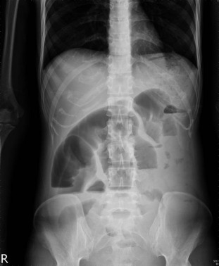
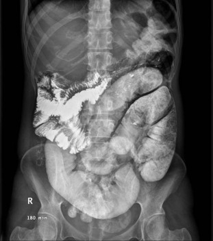
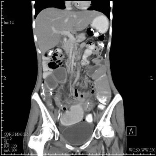
Baseline blood examinations revealed: hemoglobin 9.0 g/dL, white blood cell count 6200/mm3 , protein 3.0 g/dL, albumin 1.1 g/dL, total cholesterol 91 mg/dL, and normal renal and liver biochemistry. Plain abdominal film showed segmental dilatation of jejunal loops and intestinal obstruction was considered (Figure 1 ). A barium meal study disclosed marked dilatation of the segmental ileal bowel loops with air-fluid level and suggestion of mechanical obstruction (Figure 2 ). Abdominal computed tomography (CT) demonstrated significant dilatation of the distal jejunum to the proximal ileum, with a suspicious thickened wall in some segments probably due to inflammation (Figure 3 ).
|
|
|
Figure 1. Plain abdominal film shows segmental dilatation of jejunal loops and intestinal obstruction. |
|
|
|
Figure 2. Barium meal study discloses marked dilatation of the segmental ileal bowel loops with air-fluid level. |
|
|
|
Figure 3. Abdominal computed tomography demonstrates significant dilatation of distal jejunum to proximal ileum with suspicious thickened wall in some segments. |
What is the diagnosis?
Answer
A surgeon was consulted for operation due to mechanical obstruction from the distal jejunum to the proximal ileum. During laparotomy, the jejunum directed from the Treitz ligament 120 cm and was adhered 60 cm in length, and several engorged lymph nodes along the mesentery were found. Resection of partial jejunum was performed and pathology reported a diverticulum, 1.3 cm in greatest dimension. The mucosa was focal hemorrhage and ulceration. The surrounding tissue was hemorrhagic and necrotic. Microscopically, it showed small intestinal tissue with fistula tract formation and peritoneal adhesion to the adjacent tissue. The adjacent mucosa showed ulcer and chronic inflammation. The picture is compatible with small intestine with transmural diverticulitis. After intestinal anastomosis, the patient recovered well and was discharged.
The reported incidence of jejunoileal diverticula is 0.3–1.3% in autopsy series and 2.3% in radiographic findings [1] . Most patients with acquired jejunoileal diverticula are in the 6th –7th decade of life or older [2] . About 80% of jejunoileal diverticula occur in the jejunum [3] . This disorder usually remains asymptomatic until it presents with complications. If symptomatic, localized epigastrically or periumbilically with a bloating sensation after food intake, vomiting, diarrhea, or malabsorption are frequently seen, however, these symptoms are nonspecific. Acute complications are diverticulitis, with or without perforation. Small bowel perforation can be delayed and peritonitis develops slowly because the adjoining bowel loops make peritonitis become insignificant [1] ; [4] . The diagnosis of jejunoileal diverticula before operation was not easy. About 25% of patients could get an accurate diagnosis pre-operatively [5] .
CT is a useful tool for evaluating small bowel diverticulitis with ileus. The criteria for CT diagnosis of obstruction include discrepancy in the caliber of small-bowel loops with a definable zone of transition. Criteria for CT diagnosis of acute diverticulitis include a spectrum of changes in the pericolic fat ranging from minimal clouding to frank abscess formation [6] .
In our patient, the initial presentation was severe abdominal pain and then the symptoms shifted to bowel habit change. When a young female patient complains of alternating diarrhea and constipation combined with poor nutrition status, her history should be traced carefully.
Conflicts of interest
All authors declare no conflicts of interest.
References
- [1] W.T. Kassahun, J. Fangmann, J. Harms, M. Bartels, J. Hauss; Complicated small-bowel diverticulosis: a case report and review of the literature; World J Gastroenterol, 13 (2007), pp. 2240–2242
- [2] D.C. Chow, M. Babaian, H.L. Taubin; Jejunoileal diverticula; Gastroenterologist, 5 (1997), pp. 78–84
- [3] W.E. Longo, A.M. Vernava; Clinical implications of jejunoileal diverticular disease; Dis Colon Rectum, 35 (1992), pp. 381–388
- [4] K. Makris, G.G. Tsiotos, V. Stafyla, G.H. Sakorafas; Small intestinal nonmeckelian diverticulosis; J Clin Gastroenterol, 43 (2009), pp. 201–207
- [5] E.J. Chiu, Y.M. Shyr, C.H. Su, C.W. Wu, W.Y. Lui; Diverticular disease of the small bowel; Hepatogastroenterology, 47 (2000), pp. 181–184
- [6] A.V. Kim, G.L. Bennett, B. Perlman, A.J. Megibow; Small-bowel obstruction associated with sigmoid diverticulitis: CT Evaluation in 16 Patients; AM J Roentgenol, 170 (1998), pp. 1311–1313
Document information
Published on 15/05/17
Submitted on 15/05/17
Licence: Other
Share this document
Keywords
claim authorship
Are you one of the authors of this document?


