Summary
The use of a homemade balloon dilator and protector hood composed of condoms for assisting the removal of sharp foreign bodies lodged in the upper esophagus in difficult cases is reported. A conventional endoscopic method failed to remove two sharp bones and two press-through packages became impacted in the upper esophagus. A condom was used to make a balloon dilator that was attached to a flexible endoscope in an attempt to dilate the upper esophageal sphincter to dislodge the impacted sharp bones. This handmade condom balloon dilator succeeded in dislodging the two tightly impacted sharp bones and assisted in removing the impacted objects in the upper esophagus. Additionally, a condom was tied to the distal end of the scope to act as a protector hood to protect the esophageal mucosa when removing the sharp packages. The two impacted press-through packages were pushed into the lower esophagus or stomach and removed uneventfully using the condom protector hood. Subsequent endoscopy disclosed no relevant mucosal damage after the successful removal and the patients did well after discharge from the emergency department. In conclusion, condom-based endoscopic balloon dilatation is a simple and accessible method for assisting the endoscopic removal of tightly impacted, sharp foreign bodies in the upper esophagus. A condom can also be used as a protector hood to avoid mucosal injury when removing impacted, sharp press-through packages when a commercial protector hood is not available.
Keywords
Balloon dilator ; Condom ; Foreign body ; Impaction
Introduction
Sharp foreign body (FB) impaction in the upper esophagus is dangerous and should be addressed as soon as possible because more complications are associated with delayed removal [1] . However, it is difficult to dislodge impacted, large, sharp FBs in the upper esophagus because of the limited working space and because esophageal injury may result from attempted forced removal [2] . Here, a simple method is presented whereby condoms were used to assist flexible endoscopy for remove of sharp objects lodged in the upper esophagus in difficult cases. The accessory instruments and methods of flexible endoscopy that are used to remove sharp objects impacted in the upper esophagus are reviewed.
Case Reports
Patient 1 was a 72-year-old man and Patient 2 was a healthy 52-year-old woman, who presented to the emergency department (ED) with odynophagia after eating a fish head several hours previously. Esophagogastroduodenoscopy showed a large fish bone impacted in the upper esophagus of these two patients. The bones were firmly impacted in the bilateral esophageal wall and they could not be moved by rat-tooth forceps or by endoscopic push. Patient 3 was a 31-year-old woman and Patient 4 was a 54-year-old woman, who presented to the ED with neck pain and odynophagia within 1 hour after swallowing a press-through pack by accident. Esophagogastroduodenoscopy disclosed the large pack, with sharp and pointed edges, lodged in the upper esophagus. The packs were pushed into the lower esophagus by a scope but failed to be pulled into an overtube.
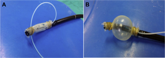
Method 1. Two layers of condoms (Taiwan Fuji, Taipei) were fastened, with a biliary cannula catheter (Olympus PR214Q, Tokyo, Japan) inside, to the distal end of the endoscope (Olympus GIF-XQ-240, Tokyo, Japan) using rubber bands to make an oral-side endoscopic balloon dilator (Fig. 1 ). The scope was intubated close to the foreign body without conscious sedation and the balloon dilator was inflated to ∼3 cm to release one end of the fish bone from the esophageal wall. The fish bone was then pushed by a scope or forceps, captured with rat-tooth forceps, pulled into the overtube, and extracted (Fig. 2 ). Subsequently, endoscopy revealed a mucosal ulcer at the impacted site in Patient 1 and Patient 2. They were discharged from the ED after observation for several hours and no complications subsequently developed.
|
|
|
Figure 1. (A) Two layers of condom and a cannula were tied to the distal end of the scope with rubber bands. (B) The balloon was inflated with air through the cannula.
|
|
|
|
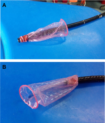
Figure 2. (A) A condom was fastened to the scope end with a rubber band to act as a protection hood. (B) The rubber ring kept the hand-made hood open when the condom slipped over the scope during withdrawal of the scope.
|
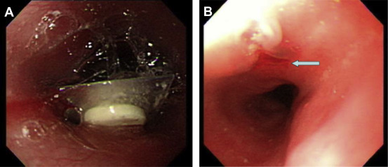
Method 2. A single condom was tied to the distal end of a scope with a rubber band, leaving a wide opening 6–8 cm away from the endoscopic lens (Fig. 3 ). A dry run was then performed in vivo to confirm that the wide opening could freely flip away from the scope to cover the captured FB when the scope was drawn through the narrow lumen. The two impacted packages were captured and were covered by the condom protector hood when removed by the scope. Patient 3 had mucosal erythema in the upper esophagus and was discharged after removal. Patient 4 was observed overnight in the ED because of mucosal ulceration at the impacted esophageal wall ( Fig. 4 ). Both patients did well after discharge.
|
|
|
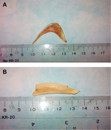
Figure 3. (A) A crescent-shaped fish bone with a sharp, pointed end and edge 3.5 cm × 1 cm was removed from Patient 1. (B) A chicken bone 3.5 cm × 1.5 cm with a pointed end was removed from Patient 2.
|
|
|
|
Figure 4. (A) A press-through-package 2 cm in largest diameter with a sharp edge impacted at the upper esophagus. (B) After endoscopic removal of the package with a condom-made protection hood, mucosal ulceration was seen at the impacted site (arrow).
|
Discussion
Although an esophageal FB is more accessible than FBs in other parts, it can cause more serious complications when the object is sharp or corrosive because the esophagus lacks a serosal layer and it is close to vital structures [3] . It is more of a struggle to remove FBs impacted in the upper esophagus because the visual field can not be secured and because the working space is limited due to the upper esophageal sphincter. Additionally, there is a higher risk of esophageal perforation in cases of inappropriate attempts to remove impacted sharp objects from the upper esophagus [2] .
Flexible endoscopy has played a major role in removing ingested FBs due to the accessibility, lower cost, popular training program, and high success rate [4] ; [5] . The accessory instruments commonly used to prevent mucosal damage when removing sharp FBs include a plastic overtube and a latex protector hood, whether homemade or commercial [6] ; [7] ; [8] . For large, sharp object impaction or deep penetration into the upper esophageal wall, a transparent cap is attached to the end of the endoscope to improve the visual field and working space. Even cap-fitted colonoscopy has been reported to separate embedded large sharp objects from the esophageal wall [9] . The oral-side balloon dilator used in esophageal varices sclerotherapy has also been used to release a sharp FB impacted in the esophagus uneventfully [2] . Even using an overtube to relax the esophageal lumen and release an impacted press-through pack has also been reported [7] .
My first two patients had large fish bones impacted in the upper esophagus. A cap-fitted scope method and an overtube dilatation method were tried to release the tight lodgments but failed. Previously, Jeen et al dilated the upper esophagus of eight patients with a commercial oral-side balloon dilator and removed each impacted FB without complications [2] . I used this handmade condom balloon to assist removing the impacted FB in the upper esophagus because a commercial balloon dilator was not available here. To avoid obstructing the airway orifice, the length of the homemade balloon dilator was adjusted to < 3 cm, given that these two impactions were located at 18 cm from the incisors. During the gradual dilatation of the balloon, the patient appeared to be well oxygenated without conscious sedation. In my limited experience, secured immediate deflation of the balloon before this procedure and oxygenation monitoring are strongly advised to avoid airway compression in case of balloon malposition.
Press-through packs have four razor-sharp edges that can perforate the esophagus when impacted or when the impacted packs are removed [10] . Most press-through packs may be removed endoscopically with the aid of a fitted cap or plastic overtube to prevent esophageal injury. Packs impacted in the esophageal wall can be released with an oral-side balloon dilator and removed using a protector hood [2] . Even a pack larger than the 15-mm overtube diameter can be removed using the overtube method by forceful pulling, bending the edge of the pack and enabling its entry into the overtube [7] . However, the overtube method failed to remove the two press-through packs in the present report because of the large size of the packs. A commercial protector hood and modified protector hood composed of a surgical latex glove were safe for removing razor blades and pointed, sharp metallic fragments [6] ; [8] . In the present cases of press-through-package impaction, I preferred to use a condom as a protector hood because of its comparable size to the package and because of its formed rubber opening ring, resisting hood collapse. This modified condom protector hood succeeded in assisting the removal of these packs without complications, and patients exhibited good tolerance. Although the application of a condom protector hood was successful and caused less discomfort during the removal of the two sharp packs, the adequacy of protection for removing sharper FBs such as a knife or razor blade should be determined in future trials, because of the ultrathin thickness of condoms (0.04–0.09 mm) compared with surgical gloves (0.29 mm) and latex hoods (2 mm: Ballard Medical).
To address difficult cases of FB impaction in the upper esophagus, a homemade condom balloon dilator is an easily accessible and safe option when other methods fail. Additionally, a modified condom protector hood may protect the mucosa when extracting the ingested sharp press-through packs if a commercial protector is not available.
Conflicts of interest
All authors declare no conflicts of interest.
References
- [1] G.M. Eisen, T.H. Baron, J.A. Dominitz, D.O. Faigel, J.L. Goldstein, J.F. Johanson, et al.; Guideline for the management of ingested foreign body; Gastrointest Endosc, 55 (2002), pp. 802–806
- [2] Y.T. Jeen, H.J. Chun, C.W. Song, S.H. Um, S.W. Lee, J.H. Choi, et al.; Endoscopic removal of sharp foreign bodies impacted in the esophagus; Endoscopy, 33 (2001), pp. 518–522
- [3] A. Chaikhouni, J.M. Kratz, F.A. Crawford; Foreign bodies of the esophagus; Am Surg, 51 (1985), pp. 173–179
- [4] D. Gmeiner, B.H.A. Von Rahden, C. Meco, J. Hutter, G. Oberascher, H.J. Stein; Flexible versus rigid endoscopy for treatment of foreign body impaction in the esophagus; Surg Endosc, 21 (2007), pp. 2026–2029
- [5] D.M. Chaves, S. Ishioka, V.N. Felix, P. Sakai, J.J. Gama-Rodrigues; Removal of a foreign body from the upper gastrointestinal tract with a flexible endoscope: a prospective study; Endoscopy, 36 (2004), pp. 887–892
- [6] G. Bertoni, R. Sssatelli, R. Conigliaro, G. Bedogni; A simple latex protector hood for safe endoscopic removal of sharp-pointed gastroesophageal foreign bodies; Gastrointest Endosc, 44 (1996), pp. 458–461
- [7] Y.S. Seo, J.J. Park, J.H. Kim, J.Y. Kim, J.E. Yeon, J.S. Kim, et al.; Removal of press-through-packs impacted in the upper esophagus using an overtube; World J Gastroenterol, 28 (2006), pp. 5909–5912
- [8] L.S. Kao, T. Nguyen, J. Dominitz, M.H.L. Teicher, D.J. Kearney; Modification of a latex glove for the safe endoscopic removal of a sharp gastric foreign body; Gastointest Endosc, 52 (2000), pp. 127–129
- [9] J.J. Hyun, H.J. Chun, B. Keum, Y.S. Seo, Y.S. Kim, Y.T. Jeen, et al.; Alternative salvage technique for removing large sharp foreign body near upper esophageal sphincter; Surg Laparosc Endosc, 22 (2012), pp. e48–e52
- [10] T. Sudo, S. Sueyoshi, H. Fujita, H. Yamana, K. Shirouzu; Esophageal perforation caused by a press through pack; Dis Esophagus, 16 (2003), pp. 169–172
Document information
Published on 15/05/17
Submitted on 15/05/17
Licence: Other
Share this document
Keywords
claim authorship
Are you one of the authors of this document?


