Summary
Expertise in enteral nutrition is an important aspect of the skill set of clinical gastroenterologists. Feeding tubes can be placed by bedside, endoscopic, fluoroscopic, and surgical methods. Nasojejunal tube placement in patients with variations in anatomy or with a history of abdominal surgery was previously a difficult problem. Today, however, as a result of developments in enteroscopy, gastroenterologists can obtain a better understanding of the small intestine. This paper reports a new technique in nasojejunal tube insertion. The feeding tube was placed in the intestine using a guide wire which accessed the jejunum with the help of single-balloon enteroscopy. Using this new skill, the placement of an enteral tube is more comfortable and safer for patients.
Keywords
Endoscopy ; Feeding tube placement ; Single-balloon enteroscopy
1. Introduction
Enteral feeding is the favored route of nutritional support, with the advantages of a decreased rate of infection, increased mucosal function, and improved immunological function. In patients with an intolerance of gastric feeding, it is recommended that a feeding tube should be placed into the duodenum or jejunum. Various methods of endoscopic placement of a nasoenteral feeding tube have been developed [1] and the standard bedside procedures often require transoral endoscopy. For two patients who had difficulty with conventional endoscopic placement of a nasoenteral feeding tube, we developed a new technique which used single-balloon enteroscopy to overcome the problem. For patients with changed anatomy, this technique can help gastroenterologists with the placement of a feeding tube.
2. Case reports
2.1. Case 1
A 71-year-old woman with a history of squamous cell carcinoma of the esophagus (cT3N0M0, stage IIA) had received an esophagectomy with esophageal reconstruction 1 year previously. This patient received nutritional support via a duodenostomy. She was admitted to hospital with aspiration pneumonia and was cared for in the intensive care unit due to acute respiratory failure. The gastroenterologists were consulted about the placement of an enteral feeding tube as a result of obstruction of the feeding tube from the duodenostomy route.
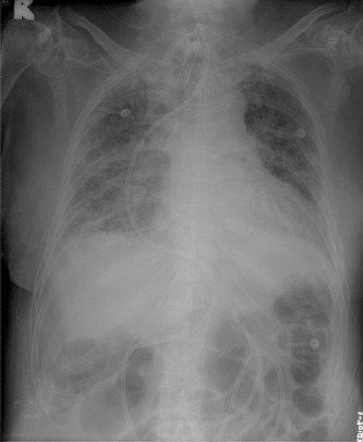
We tried to establish a feeding tube through the duodenum and a guide wire was first placed into the jejunum via single-balloon enteroscopy. The transnasal jejunal feeding tube was then pulled out from the oral cavity and a guide wire was used to access the jejunum. We held the guide wire, which passed through the lumen of the feeding tube, and slowly pushed the jejunal tube forward. Finally, we checked the position of the jejunal tube using plain film radiographs of the chest and abdomen. The procedure went smoothly and the feeding tube worked well (Fig. 1 ).
|
|
|
Figure 1. A standard 12-Fr flexible nasoenteral feeding tube is successfully placed into the jejunum. |
2.2. Case 2
A 54-year-old woman had been diagnosed 6 months previously with retroperitoneal metastatic squamous cell carcinoma (cTxNxM1) without a definitive origin. She presented on this occasion with the symptoms and signs of intestinal obstruction. An upper gastrointestinal barium study showed external compression over the third to fourth portion of the duodenum and the gastroenterologists were consulted about the placement of an enteral tube.
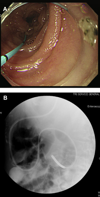
The major obstacle to directly guiding the placement of the feeding tube by endoscopy was the location of the deeper obstruction, which was only accessible by single-balloon enteroscopy. To overcome this problem, we used the same technique as in Case 1; the procedure went smoothly and the tube was successfully placed into the jejunum (Fig. 2 A and B).
|
|
|
Figure 2. (A) A guide wire is placed into the jejunum. (B) A 12-Fr feeding tube is successfully placed into the jejunum. |
3. Discussion
In our hospital, nasoenteral feeding tubes are usually advanced to the correct position with a modification of the conventional “drag and pull” technique. Suture ties (3.0-nylon) are passed through the distal end of the feeding tube, with three knots tied at 1-cm intervals on each side. With the knot grasped by forceps via the endoscope, the feeding tube can be moved with the endoscope to the appropriate position. This technique was developed by Chang et al [2] .
Other techniques have been reported for the placement of endoscopic nasoenteral feeding tubes. Wiggins and DeLegge [3] developed a modified technique with stiffening of the feeding tube via a Savary wire. Under endoscopic visualization, this tube could then be directly advanced to the correct position. Stiffening of the feeding tube can reduce the risk of pulling it out when the endoscope is withdrawn from the intestine. O'Keefe et al [4] have described a technique to place nasoenteral feeding tubes by ultrathin transnasal endoscopy. Zick et al [5] developed a technique using an indwelling feeding tube via an instrument channel in the endoscope; this may increase the ease of intervention (Table 1 ).
| Reference | Method |
|---|---|
| Chang et al [2] | 3.0-nylon suture ties were passed through the distal end of a feeding tube with three knots tied at 1-cm intervals |
| Wiggins and DeLegge [3] | Stiffening of the feeding tube via a Savary wire and then endoscopic visualization |
| O'Keefe et al [4] | Placement of nasoenteral feeding tube using ultrathin transnasal endoscopy |
| Zick et al [5] | Indwelling feeding tube via instrument channel in endoscope |
| This study | Technique mimicking nasobiliary drainage tube placement, with insertion of an enteral tube via a single balloon enteroscope |
Many routes of enteral feeding have been established, such as nasogastric tubes, nasoduodenal tubes, or nasojejunal tubes. With the advancement of single-balloon enteroscopy, nasojejunal tubes can be placed with a higher success rate. In the two patients reported here, it was difficult to place the tube into the jejunum by the method of Chang et al [2] . This was because the intestinal lumen was too long and too narrow. Using a guide wire, however, the tube could be placed through a longer distance into the desired location.
The new technique mimics the method of nasobiliary tube placement. In addition to increasing the distance of placement, the tension of the tube can be reduced by the guide wire along the greater curvature of the stomach, giving smooth advancement to the small intestine. No additional equipment is needed. This procedure can be completed quickly using only a portable endoscope, a guide wire, and a feeding tube.
Compared with percutaneous endoscopic gastrojejunostomy, more complications have been reported with nasojejunal tubes. Mucosal injury, easy aspiration, risk of acid regurgitation, esophageal stricture, and laryngospasm have been reported [6] ; [7] . Percutaneous endoscopic gastrojejunostomy is still the more favored nutritional support route, however, and, in consideration of cosmetic factors, it is more acceptable to patients. However, percutaneous endoscopic gastrojejunostomy is not indicated for all patients, particularly those who are critically ill, and portable endoscopic placement of a nasoenteral feeding tube is preferable in some patients.
In conclusion, the final decision about the methods of enteral feeding access depends on the operators experience, the patients condition, and local resources; however, single-balloon enteroscopy-assisted feeding tube placement can be one of the better choices.
Conflicts of interest
All contributing authors declare no conflicts of interest.
References
- [1] K.R. Byrne, J.C. Fang; Endoscopic placement of enteral feeding catheters; Curr Opin Gastroenterol, 22 (2006), pp. 546–550
- [2] W.K. Chang, S.A. McClave, Y.C. Chao; Simplify the technique of nasoenteric feeding tube placement with a modified suture tie; J Clin Gastroenterol, 39 (2005), pp. 47–49
- [3] T.F. Wiggins, M.H. DeLegge; Evaluation of a new technique for endoscopic nasojejunal feeding-tube placement; Gastrointest Endosc, 63 (2006), pp. 590–595
- [4] S.J. O'Keefe, W. Foody, S. Gill; Transnasal endoscopic placement of feeding tubes in the intensive care unit; J Parenter Enteral Nutr, 27 (2003), pp. 349–354
- [5] G. Zick, A. Frerichs, M. Ahrens, B. Schniewind, G. Elke, D. Schädler, et al.; A new technique for bedside placement of enteral feeding tubes: a prospective cohort study; Crit Care, 15 (2011), p. R8
- [6] N.A. Metheny, L. Schallom, D.A. Oliver, R.E. Clouse; Gastric residual volume and aspiration in critically ill patients receiving gastric feedings; Am J Crit Care, 17 (2008), pp. 512–519
- [7] S. Prabhakaran, V.A. Doraiswamy, V. Nagaraja, S. Prabhakaran, V.A. Doraiswamy, V. Nagaraja, et al.; Scand J Surg, 101 (2012), pp. 147–155
Document information
Published on 15/05/17
Submitted on 15/05/17
Licence: Other
Share this document
Keywords
claim authorship
Are you one of the authors of this document?

