Summary
We report a case of pseudolymphoma of the liver in a 49-year-old woman without an underlying disease except for liver hemangioma. A 20-mm nodule was incidentally found in segment 2 of the liver by abdominal ultrasonography during a regular follow-up of the hepatic hemangioma. After a series of radiological examinations, a left lateral sectionectomy was performed because malignant hepatic tumor could not be excluded. The patient was discharged uneventfully 7 days after the operation. The pathology examination revealed a pseudolymphoma. No recurrence of the tumor was found 5½ years after the operation. To the best of our knowledge, only 46 cases of pseudolymphoma of the liver have been reported to date. A review of the literature showed that pseudolymphomas occur predominantly in females (89.4%), usually occur as a single tumor (80.4%), are no more than 20 mm in size (90.6%), and are frequently associated with either autoimmune disease or chronic liver disease. Because an accurate diagnosis is difficult to establish, vigilant follow-up is indicated, and surgical intervention is the choice of treatment once the suspiciousness of malignancy has been raised.
Keywords
computed tomography;liver;magnetic resonance imaging;pseudolymphoma;reactive lymphoid hyperplasia
1. Introduction
Pseudolymphoma (also referred to as reactive lymphoid hyperplasia or nodular lymphoid lesion) of the liver is a rare benign nonspecific lesion characterized by a marked proliferation of polyclonal lymphocytes forming follicles with an active germinal center.1 Even though the etiology and pathogenesis of pseudolymphomas are unknown, their association with chronic infection or inflammatory process suggests their correlation with immunological response.2 In addition, their association with malignancy has been reported. The preoperative diagnosis for pseudolymphoma is difficult because noninvasive radiologic imaging findings are usually equivocal. We describe a case of pseudolymphoma and review the literature to reveal the clinicopathological characteristics of this particular disease entity.
2. Case report
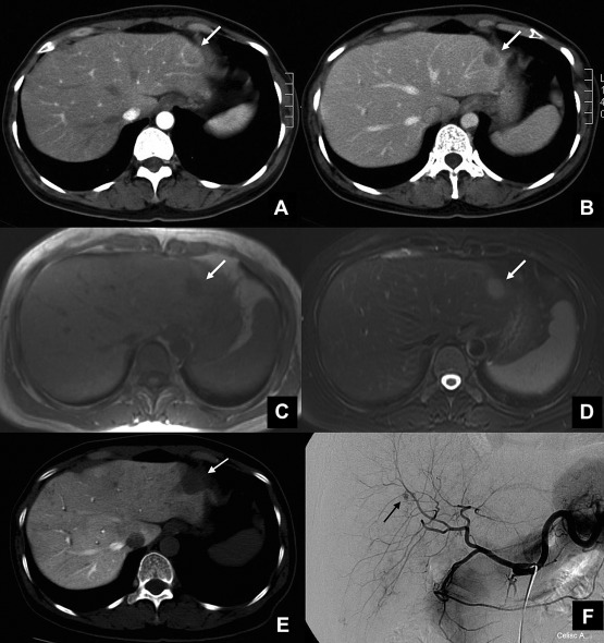
A 49-year-old woman who had a hepatic hemangioma noted since 1995 underwent an annual routine medical examination. The abdominal ultrasonography showed a hypoechoic lesion, about 20 × 16 × 14 mm in size, located in segment 2 of the liver in 2007. Laboratory tests revealed no elevation of hepatic enzymes (aspartate aminotransferase, 17 IU/L; alanine aminotransferase, 13 IU/L; alkaline phosphatase, 141 IU/L; gamma-glutamyltranspeptidase, 22 IU/L) and a normal lactate dehydrogenase level. Hepatitis B virus surface antigen and hepatitis C virus antibody were both negative. In addition, the levels of tumor markers including α-fetoprotein, carcinoembryonic antigen, and carbohydrate antigen 19-9 were all within the normal limits. The corresponding computed tomography (CT) scans revealed a relatively hypodense solid lesion with peripheral rim enhancement in arterial and portal venous phases (Fig. 1A and B). Magnetic resonance (MR) imaging showed hypointensity in axial T1-weighted images (Fig. 1C) and hyperintensity in axial T2-weighted images (Fig. 1D). Postgadolinium enhancement corresponded with the CT images. CT arterial portography demonstrated a perfusion defect in segment 2 of the liver. However, this small segment 2 tumor had no definite tumor stain on angiography study. A hypovascular tumor was impressed, and peripheral type cholangiocarcinoma, metastatic tumor, or sclerosed hemangioma should be differentiated. A left lateral sectionectomy was performed in March 2007. The patient had an uneventful postoperative course and was discharged on the 7th postoperative day.
|
|
|
Figure 1. Image findings of the lesion. (A) Abdominal computed tomography (CT) scans showed a 20-mm-diameter, slightly hypodense mass with peripheral rim enhancement in arterial phase located in segment 2. (B) Early washout of the contrast medium with retained ring enhancement was seen in the portal phase. (C) Axial magnetic resonance (MR) imaging showed a hypointense nodule on segment 2 of the liver in the T1-weighted image, and (D) the lesion became hyperintense in the T2-weighted image. (E) Arterial portography with computed tomography demonstrated a perfusion defect in segment 2 of the liver. Angiography showed a hypervascular lesion on segment 6 of the liver (arrow). (F) However, the small tumor in segment 2 of the liver on previous CT and MR had no definite tumor stain. |
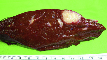
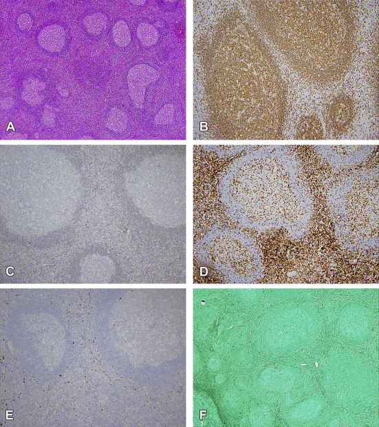
Grossly, the lesion was a well-defined, nonencapsulated, yellowish-white, and firm mass measuring 20 × 15 × 15 mm, just beneath the capsule of the liver (Fig. 2) with a satellite lesion about 3 mm in size near the previous lesion. Hematoxylin–eosin-stained histological images showed that the tumor was composed of lymphoid hyperplasia with several enlarged, irregularly shaped, well-demarcated follicles with formation of germinal centers. The lymphocytes containing round nuclei with scant cytoplasm were mainly small in size and mature in appearance with scattered medium and large cells (Fig. 3A). Tangible body macrophages were discernible. Immunohistochemical stain revealed that the follicles were composed of CD20 (+), Bcl-2 (–) B cells (Fig. 3B and C), and the interfollicular area was composed of CD3 (+) small T cells (Fig. 3D). Reactive CD30 (+) immunoblasts were evenly distributed in the interfollicular region (Fig. 3E). The EBER (Epstein–Barr virus-encoded RNA) stain was negative (Fig. 3F). The whole picture favored a diagnosis of pseudolymphoma. In addition, the satellite nodule showed similar pictures. The patient received regular follow-up, and no recurrence was found during 5½ years of follow-up.
|
|
|
Figure 2. Pathological findings of the lesion. Gross pathologic specimen revealed a well-defined, nonencapsulated, yellowish-white, and soft hepatic tumor, located just beneath the capsule in segment 2 of the liver. |
|
|
|
Figure 3. Histopathology of hepatic pseudolymphoma. Hematoxylin–eosin-stained histological images showed that the mass was composed of hyperplastic lymphoid tissue with several enlarged, irregularly shaped, well-demarcated follicles with formation of germinal centers distributed evenly in the mass. (A) The lymphocytes containing round nuclei with scant cytoplasm are mainly small in size and mature in appearance with scattered medium and large cells, ×40. Immunohistochemical stain showed that the germinal centers were composed of B lymphocytes (B) positive to CD20 antibody, ×100, (C) but negative to Bcl-2 (–) antibody, ×100. (D) The interfollicular area was composed of small T lymphocytes positive to CD3 antibody, ×100. (E) Reactive immunoblasts positive to CD30 antibody were evenly distributed in the interfollicular region, ×100. (F) However, the Epstein–Barr virus-encoded RNA stain was negative, ×40. |
3. Discussion
Pseudolymphoma was first described in the lung by Saltzstein in 19633 as a lymphocytic tumor associated with inflammation and with no evidence of systemic dissemination. Pseudolymphoma of the liver was first reported by Snover et al4 in 1981. This lymphoproliferative condition is also referred to as reactive lymphoid hyperplasia or nodular lymphoid lesion. Pseudolymphomas can be found in the gastrointestinal tract,5 orbit,6 and pancreas,7 but rarely in the liver. Based on a review of the PubMed database from 1981 to 2012 using the keywords “pseudolymphoma” and “lymphoid hyperplasia of the liver”, we found 64 lesions in 46 cases of pseudolymphoma (Table 1),1; 2; 4; 8; 9; 10; 11; 12; 13; 14; 15; 16; 17; 18; 19; 20; 21; 22; 23; 24; 25; 26; 27; 28; 29; 30; 31; 32; 33; 34; 35; 36; 37; 38; 39; 40 ; 41 and most of the reports (37 reports, 80.4%) dealt with a single tumor. Interestingly, most of the cases (69.6%) were reported in Japan. However, whether this is attributable to racial/ethnic differences or lifestyle factors such as sashimi intake was not clear. When our case was included, the average age of the patients was 56.7 years (range = 15–85, median = 58) with female predominance (F/M = 42:5, 89.3%). The average size of the tumor was 15.1 ± 10.6 mm (range, 1–60), and most of the tumors (90.6%) were no more than 20 mm. Some of the patients presented with extrahepatic autoimmune disease [Sjögrens syndrome in 1 case; autoimmune thyroiditis in 3 cases; Takayashu aortitis with antiphospholipid syndrome in 1 case; calcinosis, Raynauds phenomenon, esophageal dysmotility, sclerodactyly, telangiectasia (CREST) syndrome in 1 case—representing a total of 12.8%]. Therefore, a relationship between pseudolymphoma of the liver and immunologic dysregulation was suggested.40 Many of the patients were found to have chronic liver disease (primary biliary cirrhosis in 6 cases, viral hepatitis in 5 cases, nonalcoholic steatohepatitis in 2 cases, representing a total of 35.1%). Pseudolymphomas have been reported to develop after interferon treatment for chronic hepatitis.26 These findings support the inflammatory nature of the lesion. More importantly, many of the patients had an accompanying malignancy [colon cancer in 4 cases, gastric cancer in 3 cases, triplicate carcinomas (gastric, cecal, and colon cancer) in 1 case, renal cell carcinoma in 3 cases, ovarian cancer in 1 case, and pancreatic cancer in 1 case, representing a total of 27.7%]. Therefore, immunoreaction again carcinoma cells might relate to the development of pseudolymphoma.32 Other associated diseases include diabetes mellitus, hypertension, and diverticulitis. Interestingly, decreasing size in pseudolymphoma had been reported.23 ; 40 This phenomenon might reflect the benign behavior of this particular tumor. To date, the pathophysiology of pseudolymphoma has not been understood.
| Case | Author(s) | Age | Sex | Tumor number | Tumor size (mm) | Preoperative diagnosis | Treatment | Associated disease |
|---|---|---|---|---|---|---|---|---|
| 1 | Snover et al4 | 15 | F | 1 | N/A | Cirrhosis | N/A | Primary immunodeficiency |
| 2 | Tanabe et al9 | 30 | F | 1 | 15 | Unknown | Resection | Acute enteritis |
| 3 | Jiménez et al10 | 34 | F | 1 | 27 | Not reported | Resection | Nil |
| 4 | Willenbrock et al11 | 36 | F | 1 | 18 | N/A | N/A | Focal nodular hyperplasia of the liver, hemangioma |
| 5 | Endou et al8 | 38 | F | 1 | 18 | N/A | N/A | Pancytopenia |
| 6 | Zen et al40 | 40 | M | 1 | 20 | N/A | Resection | Chronic hepatitis B |
| 7 | Ohtsu et al12 | 42 | F | 3 | 14, 15, and smaller | HCC | Resection | Hepatitis B |
| 8 | Lin13 | 44 | F | 1 | 15 | Metastatic tumor | Resection | Colon cancer |
| 9 | Park et al14 | 46 | F | 1 | 10 | Metastatic tumor | Resection | Renal cell carcinoma, Hepatitis B |
| 10 | Nagano et al15 | 47 | F | 1 | 17 | HCC | Resection | Autoimmune thyroiditis |
| 11 | Fukuo et al34 | 47 | F | 2 | 5, 15 | HCC or inflammatory pseudotumor | Resection | Primary biliary cirrhosis |
| 12 | Okubo et al16 | 49 | F | 1 | 20 | Suspected HCC | Resection | Sjogrens syndrome |
| 13 | Mori et al17 | 49 | F | 1 | 18 | N/A | N/A | Hepatitis B |
| 14 | Shiozawa et al18 | 51 | F | 1 | 20 | N/A | N/A | Nil |
| 15 | Sharifi et al1 | 52 | F | 1 | 4 | Primary biliary cirrhosis | Transplantation | Primary biliary cirrhosis |
| 16 | Machida et al21 | 53 | F | 3 | 10, 12 and 15 | HCC | Resection | Autoimmune thyroiditis |
| 17 | Sharifi et al1 | 56 | F | 1 | 15 | Primary biliary cirrhosis | Transplantation | Primary biliary cirrhosis CREST syndrome |
| 18 | Sharifi et al1 | 56 | M | 1 | 7 | Not reported | Resection | Diverticulitis |
| 19 | Hayashi et al37 | 56 | F | 1 | 15 | Metastatic tumor or HCC | TAE, resection | Nonalcoholic fatty liver disease |
| 20 | Fujinaga et al19 | 58 | F | 1 | 15 | N/A | N/A | Hypertension |
| 21 | Nichijima et al20 | 58 | F | 1 | 12 | N/A | N/A | Hypertension, diabetes mellitus |
| 22 | Yoshikawa et al41 | 58 | F | 1 | 8 | Metastatic tumor | Resection | Gastric cancer |
| 23 | Domínguez-Pérez et al35 | 58 | F | 1 | 10 | Malignancy, premalignancy | Resection | Primary biliary cirrhosis |
| 24 | Marchetti et al38 | 58 | F | 1 | 12 | Metastatic tumor | Resection | Ovarian cancer |
| 25 | Isobe et al22 | 59 | F | 1 | 9 | Malignant tumor | Resection | Diabetes mellitus |
| 26 | Domínguez-Pérez et al35 | 59 | F | 1 | 20 | HCC | Transplantation | Advanced liver cirrhosis |
| 27 | Osame et al39 | 60 | F | Multiple | 5, 5, 6, 6, 7, 8, 10, 10, 35, 60, and several tiny ones | Metastatic tumor | Resection | Renal cell carcinoma |
| 28 | Yoshikawa et al41 | 61 | F | 1 | NA | Metastatic tumor | Resection | GIST |
| 29 | Yoshikawa et al41 | 62 | F | 1 | 4 | Metastatic tumor | Resection | Pancreatic cancer |
| 30 | Ota et al23 | 63 | F | 1 | 16 | Reactive lymphoid hyperplasia | Observation | Nil |
| 31 | Okada et al24 | 63 | F | 2 | 4, 13 | HCC | Resection | Primary biliary cirrhosis, primary aldosteronism |
| 32 | Zen et al40 | 63 | F | 2 | 5, 9 | Reactive lymphoid hyperplasia | Resection | Primary biliary cirrhosis Autoimmune thyroiditis Adrenocortical adenoma |
| 33 | Takahashi et al2 | 64 | F | 1 | 10 | HCC | Resection | Colon cancer |
| 34 | Zen et al40 | 64 | F | 2 | 10, 35 | N/A | Biopsy | Takayasu aortitis Antiphospholipid syndrome |
| 35 | Katayanagi et al25 ; 40 | 66 | F | 1 | 15 | Suspected lymphoma | Resection | Diabetes mellitus Nonalcoholic steatohepatitis |
| 36 | Tanizawa et al26 | 67 | F | 1 | 20 | HCC | Resection | Nil |
| 37 | Matsumoto et al27 | 67 | F | 1 | 12 | Reactive lymphoid hyperplasia | Ethanol injection | Hypertension |
| 38 | Kobayashi et al36 | 68 | F | 1 | 10 | Pseudotumor or metastatic tumor | Resection | Colon cancer |
| 39 | Pantanowitz et al28 | 69 | F | 1 | 17 | Metastatic tumor | Resection | Renal cell carcinoma |
| 40 | Okuhama et al29 | 70 | M | 1 | 40 | HCC | Resection | Nil |
| 41 | Maehara et al30 | 72 | F | 1 | 15 | MALT lymphoma | Resection | Nil |
| 42 | Kim et al31 | 72 | M | 1 | 17 | HCC | Resection | Gastric cancer, hepatitis C |
| 43 | Sato et al32 | 75 | F | 2 | 14, 20 | Metastatic tumor | Resection | Gastric cancer, cecal cancer, colon cancer |
| 44 | Takahashi et al2 | 77 | F | 1 | 15 | Metastatic tumor | Resection | Colon cancer |
| 45 | Zen et al40 | 81 | M | 1 | 55 | N/A | Biopsy | Cholecystolithiasis |
| 46 | Grouls33 | 85 | F | 2 | 8, 14 | N/A | Autopsy | Gastric cancer |
| 47 | Our case | 49 | F | 2 | 20, 3 | Malignant tumor | Resection | Hepatic hemangioma |
CREST = calcinosis, Raynauds phenomenon, esophageal dysmotility, sclerodactyly, telangiectasia; GIST = gastrointestinal stromal tumor; HCC = hepatocellular carcinoma; MALT = mucosa associated lymphoid tissue; N/A = not available; TAE = transarterial embolization.
Based on previously reported cases, in terms of preoperative imaging findings, most of the lesions are depicted as hypoechoic lesions on ultrasound with or without well-defined margins27 and having a low density on plain CT but variable density in contrast CT.2 ; 41 MR imaging showed nonspecific signal intensity and angiography did not show tumor stain.41 In our patient, there were no risk factors for hepatocellular carcinoma, and the liver was not cirrhotic and the α-fetoprotein level was normal. The corresponding CT and MR images showed a solitary tumor in segment 2 of the liver with peripheral enhancement. The metastatic tumor or cholangiocarcinoma should be suspected first. Common benign tumors such as hemangiomas or focal nodular hyperplasias were not likely. Because the malignancy could not be excluded, the preoperative biopsy was an essential tool to establish a final diagnosis.2 However, for fear of needle implantation of malignant tumor cells, the patient decided to receive a left lateral sectionectomy without a biopsy. Our study, consistent with literature reports, shows that it is difficult to preoperatively distinguish pseudolymphomas from malignant tumors and metastases on the basis of radiological findings.2 ; 41 In fact, accurate preoperative diagnosis proved difficult and was achieved in only three patients,23; 27 ; 40 whereas the most frequent diagnosis before operation was malignancy (80.0%) including hepatocellular carcinoma in 12 cases, metastatic tumor in 12 cases, and malignant tumor in four cases (3 of uncertain nature and 1 of lymphoma).
Histologically, pseudolymphomas consist of hyperplastic lymphoid follicles, lymphocytes, and other inflammatory cells, and an accurate diagnosis of pseudolymphoma relies on immunohistochemical analysis. Usually, immunohistochemical staining showed positive for CD3, CD4, CD8 (T markers), and CD20 and CD79a (B cell markers), which indicated the polyclonality of the tumor. However, staining for Bcl-2, IgG4, ALK, and SMA was negative.41 Furthermore, CD3-positive cells were mainly localized in the parafollicular area and CD20 immunostaining was positive for B follicles, whereas Bcl-2 staining was negative, indicating the pseudolymphomas reactive and nonneoplastic nature.38 In accordance with these findings, in our case, the immunohistochemical stain showed that the follicles were composed of CD20 (+), Bcl-2 (–) B cells, and the interfollicular area was composed of CD3 (+) small T cells. Moreover, reactive CD30 (+) immunoblasts were evenly distributed in the interfollicular region, and the EBER stain was negative.
Even though their precise etiology and pathogenesis remain unknown, pseudolymphomas have a much better prognosis than malignant lymphomas. Treatment methods for the 40 patients were recorded: resection in 32 cases (80.0%), liver transplantation in three cases, biopsy in two cases, ethanol injection in one case, autopsy in one case, and observation in one case. Ten of the 47 cases have multiple tumors (21.3%) and only one case had more than three tumors.39 Furthermore, our case was the second case associated with hepatic hemangioma,11 and the patient survived well 5½ years after the operation without tumor recurrence.
In conclusion, pseudolymphoma of the liver should be considered in the differential diagnosis of small hepatic tumors (≤ 20 mm), especially when a single hypovascular tumor is found in a female patient and the patient has no risk factors of hepatocellular carcinoma. Because it is difficult to preoperatively distinguish pseudolymphomas from malignant tumors and metastases on the basis of radiological findings, vigilant follow-up of patients is essential and surgical intervention is the choice of treatment once the suspiciousness of malignancy has been raised.
Acknowledgments
We thank Professor Kengo Yoshimitsu, Chairman of the Department of Radiology, and Dr Akinobu Osame, Faculty of Medicine, Fukuoka University, for providing unpublished data in their work. In addition, the authors thank Ms Eunice J. Yuan for secretarial assistance. Authors' contributions: conception and design of study, KLL, RHY; acquisition of data, CTY, RHY; analysis and/or interpretation of data, KLL, MCL, RHY; drafting the manuscript, CTY; revising the manuscript critically for important intellectual content, KLL, MCL, RHY; approval of the version of the manuscript to be published (the names of all authors must be listed), CTY, KLL, MCL, RHY.
References
- 1 S. Sharifi, M. Murphy, M. Loda, G.S. Pinkus, U. Khettry; Nodular lymphoid lesion of the liver: an immune-mediated disorder mimicking low-grade malignant lymphoma; Am J Surg Pathol, 23 (1999), pp. 302–308
- 2 H. Takahashi, H. Sawai, Y. Matsuo, et al.; Reactive lymphoid hyperplasia of the liver in a patient with colon cancer: report of two cases; BMC Gastroenterol, 6 (2006), p. 25
- 3 S.L. Saltzstein; Pulmonary malignant lymphomas and pseudolymphomas: classification, therapy, and prognosis; Cancer, 16 (1963), pp. 928–955
- 4 D.C. Snover, A.H. Filipovich, L.P. Dehner, W. Krivit; ‘Pseudolymphoma’. A case associated with primary immunodeficiency disease and polyglandular failure syndrome; Arch Pathol Lab Med, 105 (1981), pp. 46–49
- 5 J.J. Brooks, H.T. Enterline; Gastric pseudolymphoma. Its three subtypes and relation to lymphoma; Cancer, 51 (1983), pp. 476–486
- 6 S.J. Ryan, L.E. Zimmerman, F.M. King; Reactive lymphoid hyperplasia. An unusual form of intraocular pseudotumor; Trans Am Acad Ophthalmol Otolaryngol, 76 (1972), pp. 652–671
- 7 H. Nakashiro, O. Tokunaga, T. Watanabe, K. Ishibashi, T. Kuwaki; Localized lymphoid hyperplasia (pseudolymphoma) of the pancreas presenting with obstructive jaundice; Hum Pathol, 22 (1991), pp. 724–726
- 8 T. Endou, Y. Isobe, K. Matsunaga, et al.; A case of pseudolymphoma of the liver (in Japanese); Clinical Imagiol, 12 (1996), p. 1230
- 9 Y. Tanabe, K. Yano, Y. Yoshida, T. Sato, K. Sakai, N. Koyanagi; A resectable case of pseudlymphoma of the liver (in Japanese); Jpn J Gastroenterol, 16 (1991), p. 240
- 10 R. Jiménez, A. Beguiristain, I. Ruiz-Montesinos, et al.; Image of the month. Reactive lymphoid hyperplasia; Arch Surg, 143 (2008), pp. 805–806
- 11 K. Willenbrock, S. Kriener, S. Oeschger, M.L. Hansmann; Nodular lymphoid lesion of the liver with simultaneous focal nodular hyperplasia and hemangioma: discrimination from primary hepatic MALT-type non-Hodgkins lymphoma; Virchows Arch, 448 (2006), pp. 223–227
- 12 T. Ohtsu, Y. Sasaki, H. Tanizaki, et al.; Development of pseudolymphoma of liver following interferon-alpha therapy for chronic hepatitis B; Intern Med (1994), p. 33
- 13 E. Lin; Reactive lymphoid hyperplasia of the liver identified by FDG PET; Clin Nucl Med, 33 (2008), pp. 419–420
- 14 H.S. Park, K.Y. Jang, Y.K. Kim, B.H. Cho, W.S. Moon; Histiocyte-rich reactive lymphoid hyperplasia of the liver: unusual morphologic features; J Korean Med Sci, 23 (2008), pp. 156–160
- 15 K. Nagano, Y. Fukuda, I. Nakano, et al.; Reactive lymphoid hyperplasia of liver coexisting with chronic thyroiditis: radiographical characteristics of the disorder; J Gastroenterol Hepatol, 14 (1999), pp. 163–167
- 16 H. Okubo, H. Maekawa, K. Ogawa, et al.; Pseudolymphoma of the liver associated with Sjögrens syndrome; Scand J Rheumatol, 30 (2001), pp. 117–119
- 17 M. Mori, Y. Koga, K. Dairaku, M. Kishikawa; A case of pseudolymphoma of the liver (in Japanese); Acta Hepatol Jpn, 43 (2002), pp. 376–380
- 18 K. Shiozawa, H. Kinoshita, H. Tsuruta, et al.; A case of pseudolymphoma of the liver diagnosed before operation; Nippon Shokakibyo Gakkai Zasshi, 101 (2004), pp. 772–778
- 19 Y. Fujinaga, O. Matsui, K. Shimizu; Reactive lymphoid hyperplasia of the liver (in Japanese); Jpn J Diagn, 17 (1997), p. 586
- 20 K. Nichijima, Y. Shimizu, K. Oonishi, K. Hasebe, S. Tani, T. Hashimoto; A case of pseudolymphoma of the liver (in Japanese); Acta Hepatol Jpn, 39 (1998), pp. 23–27
- 21 T. Machida, T. Takahashi, T. Itoh, M. Hirayama, T. Morita, S. Horita; Reactive lymphoid hyperplasia of the liver: a case report and review of literature; World J Gastroenterol, 13 (2007), pp. 5403–5407
- 22 H. Isobe, S. Sakamoto, H. Sakai, et al.; Reactive lymphoid hyperplasia of the liver; J Clin Gastroenterol, 16 (1993), pp. 240–244
- 23 H. Ota, N. Isoda, F. Sunada, et al.; A case of hepatic pseudolymphoma observed without surgical intervention; Hepatol Res, 35 (2006), pp. 296–301
- 24 T. Okada, H. Mibayashi, K. Hasatani, et al.; Pseudolymphoma of the liver associated with primary biliary cirrhosis: a case report and review of literature; World J Gastroenterol, 15 (2009), pp. 4587–4592
- 25 K. Katayanagi, T. Terada, Y. Nakanuma, T. Ueno; A case of pseudolymphoma of the liver; Pathol Int, 44 (1994), pp. 704–711
- 26 T. Tanizawa, Y. Eishi, R. Kamiyama, et al.; Reactive lymphoid hyperplasia of the liver characterized by an angiofollicular pattern mimicking Castlemans disease; Pathol Int, 46 (1996), pp. 782–786
- 27 N. Matsumoto, M. Ogawa, M. Kawabata, et al.; Pseudolymphoma of the liver: sonographic findings and review of the literature; J Clin Ultrasound, 35 (2007), pp. 284–288
- 28 L. Pantanowitz, P.F. Saldinger, M.E. Kadin; Pathologic quiz case: hepatic mass in a patient with renal cell carcinoma; Arch Pathol Lab Med, 125 (2001), pp. 577–578
- 29 Y. Okuhama, T. Kenjyo, M. Oomine, T. Kaneshiro, M. Shikiya; A case of pseudolymphoma of the liver (in Japanese); Shoukakigeka, 26 (2003), pp. 1557–1562
- 30 N. Maehara, K. Chijiiwa, I. Makino, et al.; Segmentectomy for reactive lymphoid hyperplasia of the liver: report of a case; Surg Today, 36 (2006), pp. 1019–1023
- 31 S.R. Kim, Y. Hayashi, K.B. Kang, et al.; A case of pseudolymphoma of the liver with chronic hepatitis C; J Hepatol, 26 (1997), pp. 209–214
- 32 K. Sato, Y. Ueda, M. Yokoi, K. Hayashi, T. Kosaka, S. Katsuda; Reactive lymphoid hyperplasia of the liver in a patient with multiple carcinomas: a case report and brief review; J Clin Pathol, 59 (2006), pp. 990–992
- 33 V. Grouls; Pseudolymphoma (inflammatory pseudotumor) of the liver; Zentralbl Allg Pathol, 133 (1987), pp. 565–568
- 34 Y. Fukuo, T. Shibuya, Y. Fukumura, et al.; Reactive lymphoid hyperplasia of the liver associated with primary biliary cirrhosis; Med Sci Monit, 16 (2010), pp. CS81–CS86
- 35 A.D. Domínguez-Pérez, J. Castell-Monsalve; Pseudolymphoma of the liver. Contribution of double-contrast magnetic resonance imaging (in Spanish); Gastroenterol Hepatol, 33 (2010), pp. 512–516
- 36 A. Kobayashi, T. Oda, K. Fukunaga, R. Sasaki, M. Minami, N. Ohkohchi; MR imaging of reactive lymphoid hyperplasia of the liver; J Gastrointest Surg, 15 (2011), pp. 1282–1285
- 37 M. Hayashi, N. Yonetani, F. Hirokawa, et al.; An operative case of hepatic pseudolymphoma difficult to differentiate from primary hepatic marginal zone B-cell lymphoma of mucosa-associated lymphoid tissue; World J Surg Oncol, 9 (2011), p. 3
- 38 C. Marchetti, N. Manci, M. Di Maurizio, et al.; Reactive lymphoid hyperplasia of liver mimicking late ovarian cancer recurrence: case report and literature review; Int J Clin Oncol, 16 (2011), pp. 714–717
- 39 A. Osame, R. Fujimitsu, M. Ida, S. Majima, M. Takeshita, K. Yoshimitsu; Multinodular pseudolymphoma of the liver: computed tomography and magnetic resonance imaging findings; Jpn J Radiol, 29 (2011), pp. 524–527
- 40 Y. Zen, T. Fujii, Y. Nakanuma; Hepatic pseudolymphoma: a clinicopathological study of five cases and review of the literature; Mod Pathol, 23 (2010), pp. 244–250
- 41 K. Yoshikawa, M. Konisi, T. Kinoshita, et al.; Reactive lymphoid hyperplasia of the liver: literature review and 3 case reports; Hepatogastroenterology, 58 (2011), pp. 1349–1353
Document information
Published on 26/05/17
Submitted on 26/05/17
Licence: Other
Share this document
Keywords
claim authorship
Are you one of the authors of this document?


