Abstract
Background
The risk factors along with demographic and angiographic features associated with aorto-ostial atherosclerotic coronary artery disease usually differ from that of non-aorto-ostial atherosclerotic coronary artery disease.
Objectives
This study was designed to evaluate etiology of aorto-ostial atherosclerotic coronary artery disease involving left main coronary artery (LMCA), right coronary artery or both with consideration of clinical risk factors, demographic and angiographic features.
Methods
A total of 7356 angiograms over 2 years in continuation were analyzed.
Results
116 patients were found to have aorto-ostial coronary artery disease with prevalence of 1.5. A total of 95 patients who have complete data were analyzed. Mean age was 59 ± 10 years. Prevalence in males was 5.7 times greater than female. Isolated ostial LMCA was 2 times more prevalent than isolated ostial RCA. Hypertension, diabetes and smoking were the main risk factors. 34.7% of the patients had hypercholesterolemia (> 180 mg/dl) and 26.3% of the patients had hypertriglyceridemia (> 150 mg/dl). High TC/HDL (> 3.5) ratio was seen in 77.9% of the patients. When ostial LMCA group was compared with ostial RCA group hypertriglyceridemia (Odds ratio 9.8, 95% CI, 1.7–4.2, P < 0.001) and hypercholesterolemia (Odds ratio 7.05, 95% CI, 1.7–5.7, P < 0.001) emerged as independent risk factors for ostial LMCA disease.
Conclusion
Overall there is 1.5% prevalence of atherosclerotic aorto-ostial disease of coronary arteries among patients of atherosclerotic coronary artery disease and higher proportions of patients are of male sex. Hypercholesterolemia, hypertriglyceridemia and high TC/HDL ratio can be considered as risk factors for aorto-ostial atherosclerotic coronary artery disease.
Keywords
Coronary artery disease ; Risk factors ; Aorto-ostial disease
1. Introduction
Atherosclerotic coronary artery disease is one of the leading causes of morbidity and mortality world-wide.
Patients of atherosclerotic coronary artery disease, have wide variations in their age and time of presentation, gender distribution, risk factor profile, invasive and non-invasive assessment and response to different treatment modalities.
Coronary atherosclerosis could be a focal or diffuse disease. Similarly, it could be stenotic or ectatic. Why atherosclerosis affects certain regions of the coronary arteries preferentially and why clinical manifestations occur only at certain times, are still enigmas.
The spatial heterogeneity of coronary atherosclerosis could be explained on the basis of the risk factors like lipoprotein and smoking, but these have global rather than local effect on arteries.
It is well known that CAD is more common in males, as compared to females. Hypertension, diabetes, smoking and dyslipidemia are other established risk factors for CAD. Since CAD per se is more predominant in males, theoretically the same should be true for ostial CAD. However, an extensive literature review brought forward multiple studies which indicate that female sex is an independent risk factor for ostial CAD [1] and [2] . LMCA narrowing mostly occurs beyond the ostium in the mid portion or at the bifurcation, where it can extend to both major branches [3] and [4] .
The most frequent cause of LMCA stenosis, whether the ostium is affected or not, is atherosclerosis.
There is a disagreement about whether ostial LMCA disease is different from the disease in the body of the LMCA. Studies have suggested that ostial LMCA stenosis is a distinct pathology in younger and female patients with fewer risk factors for cardiovascular diseases [3] and [5] . The frequency of concomitant ostial right coronary artery (RCA) stenosis varies from 2.6% to 9% to 14% in patients with ostial LMCA stenosis [1] , [2] and [6] .
Darabian et al. suggested that risk factors such as hypertriglyceridemia and female sex are independent risk factors for ostial lesions of both LMCA and RCA [2] .
In a study of 1254 patients with coronary artery disease who underwent cardiac catheterization studies from 1975 through 1977, 114 (9%) had significant (≥ 50%) stenosis of the left main coronary artery (LMCA). Thirty-four of the 114 (29.8%) had stenosis of the LMCA ostium (2.7% patients). Unstable angina was more frequent in these patients, most of whom were in functional classes III and IV, than those with other LMCA lesions [7] .
O. Yildirimturk et al. documented co-existence of both ostial left main and right coronary artery ostial stenosis. According to them, both female predominance and coexistence of ostial LMCA and ostial RCA stenosis suggest a different pathological ground for this disease. The frequency of concomitant ostial right coronary artery (RCA) stenosis varies from 9 to 14% in patients with ostial LMCA stenosis [1] .
The incidence of CAD is unequivocally rising. Since atherosclerosis is the chief underlying etiology of ostial coronary artery disease, multi-vessel coronary artery disease with involvement of ostium is becoming more common. This study is conducted to see the change in incidence and various presentations of ostial coronary disease as well as pattern of involvement of other coronary arteries.
2. Methods
2.1. Study design
The present study was conducted in the Department of Cardiology, All India Institute of Medical Sciences, New Delhi. All patients undergoing invasive coronary angiography between July 2012 and December 2014 were screened for the presence of aorto-ostial coronary artery disease. Informed consent was taken after making the subjects aware of the purpose of the study. For patients, who underwent angiography prior to the start of study, records were extracted from in-patient hospital database and through telephonic contact with patients and OPD follow up. For patients who were analyzed prospectively, selective coronary angiography was performed by Judkins catheter via femoral route. Multiple views, including the right anterior oblique, left anterior oblique and anterior shallow caudal were recorded. In patients with ostial CAD, iv NTG was given to rule out coronary artery spasm and repeat angiography was performed. For the purpose of the study, ostial segment stenosis was defined as a proximal significant stenosis up to 3 mm from coronary origin [7] . Patients were considered to have significant ostial LMCA disease if the percent diameter stenosis exceeded 50% of the luminal diameter and ostial RCA disease if the per cent diameter stenosis exceeds 75% of the luminal diameter [7] . Of the 7356 coronary angiographies performed in the study period, 116 had significant ostial CAD (LMCA and RCA involvement). Of these, complete data was available in only 95 patients and these were included in the study.
3. Study population
The study population comprised of all patients of age above 18 years, investigated with coronary angiography between July 2012 and December 2014 in the institute. Patients with the following conditions which predispose to non-atherosclerotic disease or aorto-ostial lesion of coronaries were excluded from participation in the study: (1) History of radiotherapy, (2) Syphilis, (3) Rheumatoid arthritis, (4) Takayasu arteritis, (5) Aortic valve disease, (6) Aortic valve replacement, (7) Kawasaki disease, (8) Injury following coronary intervention, (9) Bypass grafts of CABG, (10) Restenosis after percutaneous interventions of aorto-ostial coronary lesions.
3.1. Statistical analysis
Data was collected on structured proforma and managed on Microsoft Excel spread sheet. The correctness of entries was checked and mistakes and omissions were rectified. Statistical processing and analysis were performed using SPSS version 17. Descriptive statistics were conducted by frequency tables. Continuous variables were expressed as mean ± SD. The chi-squared test and Fishers exact test were used to check the association between categorical variables.
3.2. Ethical approval and consent for study
Ethical approval was obtained from the Institute Ethics Committee and a written informed consent was obtained from the patients at the time of their enrolment.
4. Results
Total 116 patients had significant ostial disease (right or left coronary artery), out of this only 95 patients had full data that was required according to protocol. Hence the 21 patients were excluded from the study and the 95 patients with complete data were analyzed.
5. Demographic profile, risk factors, mode of presentation, echocardiographic parameters and lipid profile of the study group
Table 1 is the summary of the characteristics of the patients with atherosclerotic aorto-ostial coronary artery disease that were included in the study. The study population had mean age of 59 ± 10 years and mean BMI of 26.4 ± 4. Obesity was seen in 29 patients (30.6%) and 16 patients (16.8%) were found overweight. About 2/3rd patients were hypertensive. A substantial proportion of patients (about 44%) presented with acute coronary syndrome. NYHA Class II angina was reported by two-thirds of the patient population. Graded lipid profile of the participants has been mentioned in Table 1 . Overall, 33 patients (34.7%) had total cholesterol ≥ 180 mg/dl. 25 patients (26.3%) had triglycerides ≥ 150 mg/dl. Low HDL (< 40 mg/dl in males and < 50 mg/dl in females) was seen in 49 patients (51.6%). Serum LDL ≥ 130 mg/dl was seen in 9 patients (9.5%). Serum LDL level < 100 mg/dl was seen in 53 patients (55.8%). High LDL/HDL ratio was present in 7 patients (7.4%). High TC/HDL ratio was seen in 74 patients (77.9%).
| No. of patients (N = 95) | Percentage (%) | ||
|---|---|---|---|
| Age wise distribution | <55years | 32 | 34.0 |
| ≥55years | 63 | 66.0 | |
| Sex wise distribution | Male | 81 | 85.0 |
| Female | 14 | 15.0 | |
| BMI (kg/m2) | <25 | 50 | 52.6 |
| 25–29.9 | 16 | 16.8 | |
| ≥30 | 29 | 30.6 | |
| Diabetes | 35 | 36.8 | |
| Hypertension | 60 | 63.2 | |
| Smoking | 37 | 38.9 | |
| Positive family H/O CAD | 4 | 4.3 | |
| STEMI | 10 | 10.6 | |
| NSTEMI | 31 | 32.6 | |
| Non-ACS | 54 | 56.8 | |
| Angina NYHA Class II | 75 | 78.9 | |
| Angina NYHA Class III | 19 | 20.0 | |
| Angina NYHA Class IV | 1 | 1.1 | |
| LV EF<30% | 5 | 5.5 | |
| LV EF 30–45% | 20 | 21.1 | |
| LV EF 46–55% | 30 | 31.5 | |
| LV EF>55% | 40 | 42.1 | |
| Total cholesterol | ≥180mg/dl | 33 | 34.7 |
| <180mg/dl | 62 | 65.3 | |
| Triglycerides | ≥150mg/dl | 25 | 26.3 |
| <150mg/dl | 70 | 73.7 | |
| HDL | Abnormal | 49 | 51.6 |
| Normal | 46 | 48.4 | |
| LDL | <100mg/dl | 53 | 55.8 |
| 100–129mg/dl | 33 | 34.7 | |
| >130mg/dl | 9 | 9.5 | |
| LDL/HDL | ≥3 | 7 | 7.4 |
| <3 | 88 | 92.6 | |
| TC/HDL | <3.5 | 21 | 22.1 |
| ≥3.5 | 74 | 77.9 |
5.1. Pattern of vessel involvement
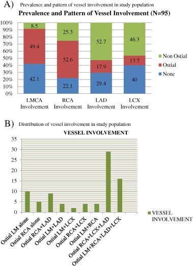
LMCA was diseased in 55 patients (57.9%) out of which 47 patients had significant ostial disease. 40 patients (42.1%) had normal LMCA. RCA was diseased in 74 patients (77.9%) out of which 50 patients had significant ostial disease. 21 patients (22.1%) had normal RCA. Isolated ostial LMCA was involved in 10 patients. Isolated ostial RCA was involved in 5 patients. One patient had involvement of both ostial RCA and ostial LMCA without involvement of distal vessels. Ostial LMCA was associated with diseased LAD in 4 patients, diseased LCX in 2 patients and diseased non-ostial RCA in 4 patients. All vessels (LMCA, RCA, LAD and LCX) were involved together in 16 patients. The most common pattern of coronary vessel involvement was ostial RCA with simultaneous LCX and LAD disease and this was seen in 29 patients. Ostial RCA with LAD disease was seen in 9 patients and ostial RCA with LCX disease was seen in 4 patients (Table 2 , Fig. 1 A and B).
| No. of patients N = 95 | Percentage (%) | ||
|---|---|---|---|
| LMCA involvement | |||
| Present | Ostial | 47 | 49.4 |
| Non-ostial | 8 | 8.5 | |
| Absent | 40 | 42.1 | |
| RCA involvement | |||
| Present | Ostial | 50 | 52.6 |
| Non-ostial | 24 | 25.3 | |
| Absent | 21 | 22.1 | |
| LAD involvement | |||
| Present | Ostial | 17 | 17.9 |
| Non-ostial | 50 | 52.7 | |
| Absent | 28 | 29.4 | |
| LCx involvement | |||
| Present | Ostial | 13 | 13.7 |
| Non-ostial | 44 | 46.3 | |
| Absent | 38 | 40.4 | |
|
|
|
Fig. 1. A. Prevalence and pattern of vessel involvement in study population. B. Distribution of vessel involvement in study population. |
5.2. Demographics of study group according to individual vessel involvement
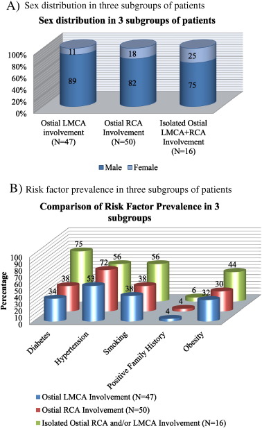
With ostial LMCA disease, 42 (89%) patients were males. The mean age of patients with LMCA disease was 57 ± 11 years. 16 (34%) were diabetic, 25 (53%) were hypertensive and 18 (38%) were smokers. 15 patients (32%) were obese. (Table 3 , Fig. 2 A and B). In the sub group of patients with ostial RCA disease, 41 (82%) patients were males. The mean age was 60 ± 9 years. 36 (72%) were hypertensive and 19 (38%) were diabetic. 19 (38%) were smokers while 15 (30%) were obese (Table 3 , Fig. 2 A and B). In the subgroup of patients with isolated RCA or LMCA disease, 12 (75%) were males. The mean age of this subgroup was 59.2 ± 5 years. 12 (75%) were diabetic and 4 (25%) were hypertensive. 9 (56%) were smokers and 7 (44%) were obese (Table 3 ,Fig. 2 A and B).
| Ostial LMCA | Ostial RCA | Isolated ostial LMCA or RCA | ||||
|---|---|---|---|---|---|---|
| N = 47 | % | N = 50 | % | N = 16 | % | |
| Male | 42 | 89 | 41 | 82 | 12 | 75 |
| Female | 5 | 11 | 9 | 18 | 4 | 25 |
| Diabetes | 16 | 34 | 19 | 38 | 12 | 75 |
| Hypertension | 25 | 53 | 36 | 72 | 9 | 56 |
| Smoking | 18 | 38 | 19 | 38 | 9 | 56 |
| Family history | 2 | 4 | 2 | 4 | 1 | 6 |
| Obesity | 15 | 32 | 15 | 30 | 7 | 44 |
|
|
|
Fig. 2. A. Sex distribution in three subgroups of patients. B. Risk factor prevalence in three subgroups of patients. |
5.3. Clinical presentation according to individual vessel involvement
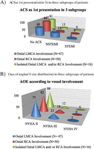
28 (60%) patients in ostial LMCA group, 48 (84%) patients in ostial RCA group and 13 (81%) patients in isolated ostial LMCA or RCA group do not have acute coronary syndrome as their first clinical presentation. Table 4 and Fig. 3 A and B are providing cumulative representation of clinical presentation according to individual vessel involvement in each of the three groups of patients.
| Ostial LMCA | Ostial RCA | Isolated ostial LMCA or RCA | |||||
|---|---|---|---|---|---|---|---|
| N = 47 | % | N = 50 | % | N = 16 | % | ||
| AOE | NYHA II | 35 | 75 | 41 | 82 | 14 | 88 |
| NYHA III | 11 | 23 | 9 | 18 | 2 | 12 | |
| NYHA IV | 1 | 2 | 0 | 0 | 0 | 0 | |
| ACS | None | 28 | 60 | 42 | 84 | 13 | 81 |
| NSTEMI | 16 | 34 | 6 | 12 | 2 | 13 | |
| STEMI | 3 | 6 | 2 | 4 | 1 | 6 | |
|
|
|
Fig. 3. A. ACS as 1st presentation (in %) in three subgroups of patients. B. Class of angina (% vice distribution) in three subgroups of patients. |
5.4. Lipid profiles according to individual vessel involvement
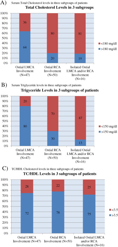
Hypercholestrolemia (total cholesterol ≥ 180 mg/dl) and hypertriglyceridemia (TG ≥ 150 mg/dl) were mainly observed in ostial LMCA group and not in other two groups (Table 5 and Fig. 4 A, B). It was observed that serum hypertriglyceridemia and hypercholesterolemia were significantly more common in patients with ostial LMCA disease than in ostial RCA disease (P < 0.001). High TC/HDL ratio (≥ 3.5) was seen in 34 (72%) patients in ostial LMCA group, 39 (78%) patients in ostial RCA group and 12 (75%) patients in isolated ostial LMCA or RCA group (Table 5 and Fig. 4 C).
| Ostial LMCA | Ostial RCA | Isolated ostial LMCA or RCA | |||||
|---|---|---|---|---|---|---|---|
| N = 47 | % | N = 50 | % | N = 16 | % | ||
| Total cholesterol | ≥180mg/dl | 30 | 64 | 10 | 20 | 3 | 19 |
| <180mg/dl | 17 | 36 | 40 | 80 | 13 | 81 | |
| Triglycerides | ≥150mg/dl | 38 | 80 | 15 | 30 | 2 | 13 |
| <150mg/dl | 9 | 20 | 35 | 70 | 14 | 87 | |
| HDL | Abnormal | 19 | 40 | 29 | 58 | 8 | 50 |
| Normal | 28 | 60 | 21 | 42 | 8 | 50 | |
| LDL | <100mg/dl | 25 | 53 | 29 | 58 | 10 | 63 |
| 100–129mg/dl | 20 | 43 | 14 | 28 | 6 | 36 | |
| ≥130mg/dl | 2 | 4 | 7 | 14 | 0 | 0 | |
| LDL/HDL | <3 | 44 | 94 | 47 | 94 | 15 | 94 |
| ≥3 | 3 | 6 | 3 | 6 | 1 | 6 | |
| TC/HDL | <3.5 | 13 | 28 | 11 | 22 | 4 | 25 |
| ≥3.5 | 34 | 72 | 39 | 78 | 12 | 75 | |
|
|
|
Fig. 4. A. Serum total cholesterol levels in three subgroups of patients. B. Serum triglyceride levels in three subgroups of patients. C. TC/HDL cholesterol levels in three subgroups of patients. |
6. Discussion
According to the projections for 2020, cardiovascular disease will remain the leading cause of death and disability in industrial countries [8] . Coronary artery disease (CAD) currently accounts for very high mortality rates in developing countries. Among patients with CAD, it is widely accepted that significant LMCA disease is associated with an increased cardiac mortality; in addition, it is the most prognostically important single lesion involving the coronary arteries. Ostial lesions presenting as an obstructive disease proximal to the bifurcation of the main stem into the LAD and LCX jeopardize all but the inferior and posterior surfaces of the left ventricle. Ostial left main coronary artery disease is frequently accompanied by the concomitant involvement of 1 or more of other epicardial vessels [9] .
The risk of CAD is associated with the extent and severity of atherosclerosis in adults. The concept that CAD can be prevented has increasingly become a driving force in cardiovascular medicine. At the core of primary prevention lies the concept of risk assessment. A general notion has evolved that the intensity of preventive efforts should be adjusted to a patients risk for developing CHD, that is, the higher the risk, the more aggressive the intervention should be.
In the current study, of the 7356 coronary angiographies performed in the said period, 116 patient had significant ostial CAD. The prevalence of ostial coronary artery disease was 1.5%. In a similar study conducted by Darabian et al. in 2008, the prevalence of significant coronary ostial disease was 2.6%. They defined significant LMCA and RCA as 50% luminal diameter stenosis [2] . In our study, the significant LMCA ostial disease is a decrease of > 50% luminal diameter and significant RCA ostial disease is a reduction of > 70% luminal diameter as per generally accepted definition. Therefore prevalence in our study may be lower due to strict criteria used that may differ from the generally accepted definition of ostial stenosis of RCA. In another study conducted by Yildirimturk et al., 87 patients had significant ostial left main coronary or significant right coronary artery disease in 2898 coronary angiographies with prevalence of 3.0% [1] .
An observation of a few studies which have studied ostial CAD has been summarized in Table 6 for comparison and contrast with present study.
| Darabian et al. 2008 [2] | Mahajan et al. [10] | Yildirimturk et al. (2011) [1] | Present study (Verma et al) | ||||||||
|---|---|---|---|---|---|---|---|---|---|---|---|
| Ostial LMCA | Ostial RCA | Ostial LMCA + RCA | Ostial LMCA | Ostial LMCA | Ostial RCA | Ostial LMCA + RCA | Ostial LMCA | Ostial RCA | Ostial LMCA + RCA | ||
| No of patients | 66 | 65 | 36 | 46 | 53 | 19 | 15 | 47 | 50 | 16 | |
| Mean age (years) | 62.36±7.8 | 63.3±8.6 | 62.64±7.94 | 65±13 | 64±11 | 66±11.9 | 67±9.6 | 57±11 | 60±9 | 59.2±5 | |
| Sex | Male | 59.1% (39) | 58.5% (38) | 52.8% (19) | 48% (22) | 84.9% (45) | 84.2% (16) | 46.7% (7) | 89% (42) | 82% (41) | 75% (12) |
| Female | 40.9% (27) | 41.5% (27) | 47.2% (17) | 52% (24) | 15.1% (8) | 15.8% (3) | 53.3% (8) | 11% (5) | 18% (9) | 25% (4) | |
| Hypercholesterolemia | – | – | – | 65% (30) | 48% (24) | 42.1% (8) | 64.3% (9) | 64% (30) | 20% (10) | 19% (3) | |
| Hypertension | 63.1% (41) | 60.9% (39) | 65.7% (23) | 72% (33) | 64% (32) | 63.2% (12) | 85.7% (12) | 53% (25) | 72% (36) | 56% (9) | |
| Diabetes | 37.5% (24) | 30.6% (19) | 35.3% (12) | 43% (20) | 38% (19) | 47.4% (9) | 42.9% (6) | 34% (16) | 38% (19) | 75% (12) | |
| Smoking | 40.9% (27) | 43% (28) | 48.5 (17) | 26% (12) | 44% (22) | 26% (5) | 21% (3) | 38% (18) | 38% (19) | 56% (9) | |
| Family history | 21.9% (14) | 27% (17) | 28.6% (10) | 39% (18) | 14% (7) | 10.5% (2) | 14.3% (2) | 4% (2) | 4% (2) | 6% (1) | |
| Obesity | – | – | – | – | 14% (7) | 26.3% (5) | 21.4% (3) | 32% (15) | 30% (15) | 44% (7) | |
| ACS | 46.1% (47) | 61.6% (40) | 53% (20) | – | – | – | – | 40% (19) | 16% (8) | 19% (3) | |
In our study, 95 patients with significant ostial coronary disease (left main and right) were selected. Mean age was 59 ± 10 years. Prevalence in males was 5.7 times greater than female (85% vs. 15%). When patients were subgrouped in ostial LMCA and ostial RCA disease, a similar pattern of male predominance was seen in both groups. Ostial LMCA was 8 times more prevalent and ostial RCA was 4 times more prevalent in males than females. When isolated ostial LMCA and ostial RCA disease group was considered, male prevalence was three times that of female (12 Vs 4, Table 3 ). These findings contradict finding in similar studies by Darabian et al. [2] and Yildirimturk et al. [1] which have observed greater female predominance in ostial CAD. In the study by Mahajan et al., there was a trend suggestive of a higher incidence of ostial lesions among women (63% vs. 31%; P = .06) [10] . Barner et al. reported that 43.5% of their left ostial group and 12% of their left main group were women (P <.005) [11] . Sasaguri et al. noticed that 4 patients out of 5 cases (80%) in their ostial stenosis group were women, but there were only 10 women (9%) in the LMCA group [3] . Yamanaka and Hobbs reported that women were predominant among subjects with stenosis of 1 or both coronary ostial (64%) [6] . Miller et al. [12] detected 5 (0.12%) women with isolated ostial Left main coronary artery disease among 4000 patients with CAD, and Welch et al. [13] reported 10 (0.01%) women with isolated left main lesion among 1000 women in their study. However, these studies do not explain this higher prevalence of ostial CAD in females despite predominance of atherosclerotic risk factors in males.
Prevalence of ostial CAD in the elderly (age ≥ 55 years) was twice the number of young patients (age < 55 years). There was no significant age difference between ostial LMCA group and ostial RCA group in our study.
Of the 95 patients, 16 patients had isolated significant ostial disease without involvement of distal part of coronary artery. Isolated ostial LMCA was involved in 10 patients. Isolated ostial RCA was seen in 5 patients. Ostial RCA disease was more common than ostial LMCA disease. This is similar to the findings by Rissanen et al. [14] .One patient had involvement of both ostial RCA and ostial LMCA without involvement of distal vessels. Ostial LMCA was associated with diseased LAD in 4 patients, diseased LCX in 2 patients and diseased non-ostial RCA in 4 patients. All vessels (LMCA, RCA, LAD and LCX) were involved concomitantly in 16 patients.
The most common pattern of coronary vessel involvement was ostial RCA with simultaneous LCX and LAD disease and this was seen in 29 patients. Ostial RCA with LAD disease was seen in 9 patients and ostial RCA with LCX disease was seen in 4 patients. This signifies that atherosclerosis is a diffuse disease and involvement of multiple segment of coronary disease is common.
Among risk factors, hypertension emerged as the chief risk factor and was present in 63.2% of the study group. Similar results are seen in studies by Darabian et al. [2] , Yildirimturk et al. [1] and Mahajan et al. [10] (Table 6 ). In our study, diabetes was present in 36.8% and smoking 39.6% patients. This statistics are similar to the findings by Mahajan et al. [10] and Yildirimturk et al. [1] . The prevalence of obesity in the study group was 30.6% of the patients. Significant family history for CAD was present in only 4% of our patients. This does not correlate with the higher prevalence of positive family history that was seen in other studies [1] , [2] and [10] .
In our study, there was no significant difference in terms of risk factors like diabetes, hypertension, smoking, family history, obesity, HDL, LDL, LDL/HDL, total cholesterol/HDL in ostial LMCA, ostial RCA and isolated ostial LMCA and RCA stenosis group.
The relationship between lipid profile and pattern of disease was studied diligently. 33 patients (34.7%) had hypercholesterolemia (≥ 180 mg/dl). 25 patients (26.3%) had hypertriglyceridemia (≥ 150 mg/dl). Low HDL (< 40 mg/dl in males and < 50 mg/dl in females) was seen in 49 patients (51.6%). Serum LDL ≥ 130 mg/dl was seen in 9 patients (9.5%). Serum LDL level ≤ 100 mg/dl was seen in 53 patients (55.8%). High LDL/HDL ratio was seen in 7 patients (7.4%). High TC/HDL ratio was seen in 74 patients (77.9%).
When subgroup analysis was done for ostial left main coronary artery disease 64% had hypercholesterolemia (≥ 180 mg/dl) and 80% had hypertriglyceridemia (≥ 150 mg/dl). Low HDL ( <40 mg/dl in males and <50 mg/dl in females) was seen in 19 patients (40%). Serum LDL ≥ 130 mg/dl was seen in 2 patients (4%). In sub group analysis of patients with ostial RCA involvement, 8% had hypercholesterolemia ( >180 mg/dl). 30% had hypertriglyceridemia (> 150 mg/dl).
When ostial LMCA group was compared with ostial RCA group hypertriglyceridemia (Odds ratio 9.8, 95% CI, 1.7–4.2, P < 0.001) and hypercholesterolemia (Odds ratio 7.05, 95% CI, 1.7–5.7, P < 0.001) emerged as independent risk factors for ostial LMCA disease.
42% patients had acute coronary syndrome (ACS) as first clinical event of presentation, out of which NSTEMI was found in 32.6% and STEMI was found in 10.6% of the patients. ACS was significantly higher in ostial left main subgroup compared to ostial RCA subgroup, (P < 0.05) while there was no significant difference in severity on angina in the two sub groups.
Ostial coronary disease is one of the manifestations of atherosclerosis and atherosclerosis incidence is higher in male. In our study frequency of male is higher than female similar to non-ostial coronary atherosclerotic disease. Ostial coronary disease is influenced by hemodynamic at the ostium as there is increased turbulence which may lead to endothelial dysfunction, endothelial injury and plaque deposition that lead to atherosclerotic stenosis. Different studies that have mentioned female preponderance to ostial coronary artery disease have not mentioned a likely cause for this observation. The study has got limitation as the sample size is small. We need to evaluate risk factors other than diabetes, hypertension, smoking, family history and lipid profile also.
7. Conclusion
We concluded that prevalence of significant ostial coronary artery disease in this study was 1.5% and higher proportion of patient was of male sex. Hypertriglyceridemia and hypercholesterolemia can be considered as risk factors for ostial left main coronary artery disease. Prevalence of ACS was higher in ostial left coronary artery disease group compared to RCA subgroup. There was no difference in terms of risk factors other than hypertriglyceridemia and hypercholestrolemia in ostial LMCA and ostial RCA group.
Conflict of interest
The authors report no relationships that could be construed as a conflict of interest.
References
- [1] O. Yildirimturk, M. Cansel, R. Erdim, E. Ozen, I.C.C. Demiroglu, V. Aytekin; Coexistence of left main and right coronary artery ostial stenosis: demographic and angiographic features; Int. J. Angiol., 20 (1) (Mar 2011), pp. 33–38
- [2] S. Darabian, A.R. Amirzadegan, H. Sadeghian, S. Sadeghian, A. Abbasi, M. Raeesi; Ostial lesions of left main and right coronary arteries: demographic and angiographic features; Angiology, 59 (2008), pp. 682–687
- [3] S. Sasaguri, Y. Honda, T. Kanou; Isolated coronary ostial stenosis compared with left main trunk disease; Jpn. Circ. J., 55 (1991), pp. 1187–1191
- [4] C.L. Pritchard, J.G. Mudd, H.B. Barner; Coronary ostial stenosis; Circulation, 52 (1975), pp. 46–48
- [5] K.K. Koh, H.K. Hwang, P.G. Kim, et al.; Isolated left main coronary ostial stenosis in oriental people: operative, histopathologic and clinical findings in six patients; J. Am. Coll. Cardiol., 21 (1993), pp. 369–373
- [6] O. Yamanaka, R.E. Hobbs; Solitary ostial coronary artery stenosis; Jpn. Circ. J., 57 (1993), pp. 404–410
- [7] B.L. Salem; Left main coronary artery ostial stenosis: clinical markers, angiographic recognition and distinction from left main disease; Catheter. Cardiovasc. Diagn., 5 (2) (1979), pp. 125–134
- [8] C.J. Murray, A.D. Lopez; Alternative projections of mortality and disability by cause 1990–2020: global burden of disease study; Lancet, 349 (1997), pp. 1498–1504
- [9] B.H. Bulkley, W.C. Roberts; Atherosclerotic narrowing of the left main coronary artery: a necropsy analysis of 152 patients with fatal coronary heart disease and varying degrees of left main narrowing; Circulation, 53 (1976), pp. 823–828
- [10] N. Mahajan, M.B. Hollander, et al.; Isolated and significant LMCA disease, demographic, hemodynamic and angiographic features; Angiology, 57 (2006), pp. 464–477
- [11] Barner HB, Reese J, Standeven J, McBride LR, Pennington DG, Willman VL. Left coronary ostial stenosis: comparison with left main coronary artery stenosis. Ann. Thorac. Surg.; 47(2):293–6.
- [12] G.A. Miller, M. Honey, H. El-Sayed; Isolated coronary ostial stenosis; Catheter. Cardiovasc. Diagn., 12 (1986), pp. 30–33
- [13] C.C. Welch, W.L. Proudfit, W.C. Sheldon; Coronary arteriographic findings in 1000 women under age 50; Am. J. Cardiol., 35 (2) (1975 Feb), pp. 211–215
- [14] R. Viljo; Occurrence of coronary ostial stenosis in a necropsy series of myocardial sudden death and violent death; BHJ, 37 (2) (1975), pp. 182–191
Document information
Published on 19/07/16
Licence: CC BY-NC-SA license
Share this document
Keywords
claim authorship
Are you one of the authors of this document?


