Question
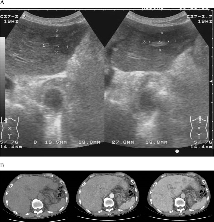
An 86-year-old man presented with intermittent fever for 1 month. He had dull abdominal pain in the right upper quadrant for 2 weeks. The patient denied a productive cough, chest pain, and dysuria. He had no recent travel history and no body weight loss. He had no underlying systemic disease, such as diabetes mellitus. Physical examinations were not remarkable. Laboratory data revealed leukocytosis (white blood cells: 21.34 × 10³ /μL, neutrophils: 83.6%). Aspartate and alanine aminotransferase levels were 23 IU/L and 19 IU/L (normal range: < 31 IU/L), respectively, and total bilirubin was 0.3 mg/dL (normal range: 0.2–1.5 mg/dL). His alkaline phosphatase was 117 IU/L (normal range: 40–129 IU/L). Hepatitis B surface antigen and antihepatitis C viral antibody were nonreactive, and tumor markers (α-fetoprotein, carcinoembryonic antigen, prostate-specific antigen, and cancer antigen 19-9) were within normal limits. An initial chest radiograph showed no abnormalities. Because of sustained fever, abdominal ultrasonography (Fig. 1 A) and computed tomography (Fig. 1 B, from left to right: noncontrast, arterial phase, and delayed phase) were performed. What is your impression?
- Hepatic bacterial abscess
- Hepatic amebiasis
- Hepatic tuberculosis
- Hepatocellular carcinoma
|
|
|
Figure 1. (A) Abdominal ultrasonography of the liver (left: right lateral lobe; right: left medical lobe); (B) computed tomography of the abdomen (from left to right: noncontrast image, arterial phase, and delayed phase of postcontrast images). |
Answer: (3)
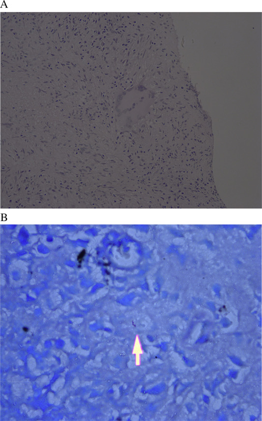
The patient still had a fever after 1 week of therapy with intravenous ceftriaxone 1 g/12 hours. Echo-guided liver aspiration and biopsy were performed. Microscopically, granulomatous inflammation with caseous necrosis accompanied by granulomas and Langhans type giant cells (Fig. 2 A) was seen. Acid-fast stain showed rare rod-like organisms (Fig. 2 B, arrow). The sputum acid-fast stain and mycobacteria culture were negative. The result of anti-human immunodeficiency virus was nonreactive. Antituberculosis medications, including rifampicin 600 mg, isoniazid 300 mg, ethambutol 800 mg, and pyrazinamide 1500 mg/day, were prescribed. The patient had no fever 1 week after taking the medications. Serial follow-up abdominal ultrasonography showed decreases in the size and number of hepatic tumors.
|
|
|
Figure 2. (A) Microscopically, granulomatous inflammation with caseous necrosis accompanied by granulomas and Langhans type giant cells is seen in the liver tissue; (B) acid-fast staining shows rod-like organisms. |
Take home message
Hepatic tuberculosis can be separated into miliary and local types by Rolleston’s classification. Primary tuberculous liver abscesses are uncommon in Taiwan nowadays. The disease should be considered in febrile patients with multiple hepatic tumors. The differential diagnosis includes primary or metastatic hypovascular carcinoma of the liver, and amebic and bacterial liver abscesses.
Conflicts of interest
All authors declare no conflicts of interest.
Document information
Published on 15/05/17
Submitted on 15/05/17
Licence: Other
Share this document
Keywords
claim authorship
Are you one of the authors of this document?

