Abstract
An 83-year-old high-risk gentleman diagnosed with severe symptomatic aortic stenosis was scheduled for TAVR. A 31 mm CoreValve was implanted but severe paravalvular leak was noted. A valve-in-valve procedure was performed. However, the valve frame was partially dislodged into de ascending aorta. We report our strategy to solve this severe leak after a failed valve-in-valve procedure.
Keywords
TAVI;Complications
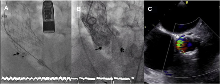
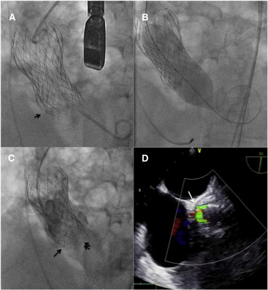
An 83-year-old high-risk gentleman diagnosed with severe symptomatic aortic stenosis was scheduled for TAVR. A CT scan showed severe bulky calcification of aortic leaflets, mainly in the non-coronary sinus. Annulus measurements were compatible with a 31 mm CoreValve™ (Medtronic, Minneapolis, Minnesota). After the full release of the valve, we observed that the frame had popped out and it was partially located above the leaflets (Fig. 1A). Severe paravalvular regurgitation (PVR) was noted (Fig. 1B and C). The decision was to proceed with a new 31 mm CoreValve, valve-in-valve technique. The second valve was deployed in a deeper position (6–8 mm) to ensure full coverage of the leak. The deployment was correct, however still severe PVR was noted (Fig. 2A). We decide to perform a balloon postdilatation with a NuCLEUS™ (NuMED Inc.) 28 mm balloon to improve the expansion of the valve structure. Interestingly, after the inflation, we realized that again the valve frame had slipped in the bulky calcification and the valve was partially dislodged (Fig. 2B, C and D). After considering different alternatives to solve the PVR after a valve-in-valve procedure, we finally decided to implant a third valve. Subsequently, we deployed a 29 mm CoreValve deep in the left ventricular outflow tract (8–10 mm), and after complete release, a new angiogram and TEE monitoring showed complete disappearance of the PVR (Fig. 3A and B). A CT scan at 1-year follow-up showed the position of valve frames and the patency of both coronary arteries (Fig. 3C and D).
|
|
|
Fig. 1. (A) Bulky calcification in non-coronary sinus (asterisk) where de valve frame is deformed. (B) Partial dislodgement of the TAV (arrow) and presence of severe aortic regurgitation (double arrow). (C) TEE monitoring showing severe paravalvular regurgitation located in non-coronary sinus and occupying almost 50% of the circumference. |
|
|
|
Fig. 2. (A) Second TAV implanted below the previous one. We can observe that the lower part is below the calcium landmark (arrow), but the valve frame is not fully expanded. (B) Balloon postdilatation trying to fully expand the valve frame. (C) After postdilatation, the valve slipped again in the severe calcification and was again partially dislodged (arrow) leading to a severe regurgitation (double arrow). (D) Severe PVL by TEE monitoring. |
|
|
|
Fig. 3. (A) Implant of third TAV far below the previous ones (arrow). (B) Only traces of aortic regurgitation were seen in TEE imaging. (C) CT scan performed 1 year after the procedure demonstrating the three valves in place (asterisk, arrow and double arrow). (D) A 3D CT scan rendering reconstruction with the three valves and demonstrating the patency of both coronary arteries. |
PVR is a major concern after TAVI since it is linked with an impaired prognosis [1]. When severe PVR is noted during the procedure usually balloon postdilatation or valve-in-valve procedures are the default treatment strategies[2]. However, the solution of a PVR after a valve-in-valve is challenging. This case is, to the best of our knowledge, the first to report a double valve-in-valve technique. We proved that a third valve implantation is feasible and may solve the complication, leading to persistent optimal results at one year of follow-up.
Disclosures
No conflict of interest to disclose.
References
- [1] M. Gilard, H. Eltchaninoff, B. Iung, et al.; Registry of transcatheter aortic-valve implantation in high-risk patients; N Engl J Med, 366 (2012), pp. 1705–1715
- [2] J.M. Sinning, M. Vasa-Nicotera, D. Chin, et al.; Evaluation and management of paravalvular aortic regurgitation after transcatheter aortic valve replacement; J Am Coll Cardiol, 62 (2013), pp. 11–20
Document information
Published on 19/05/17
Submitted on 19/05/17
Licence: Other
Share this document
Keywords
claim authorship
Are you one of the authors of this document?


