Abstract
Considering the increase in consumption of Cannabis sativa and the use of the compound β- carotene (BC) as supplement, we investigated potential changes in the chemical and biological proprieties of BC after exposure to C. sativa smoke (CSS). Our results showed that the BC exposed to CSS underwent 98.8% degradation and suffered loss of its antiradical activity. The major degradation products identified were 3-hydroxy-2,4,4-trimethylpentyl)2-methylpropanoate and (2-ethyl-3-hydroxyhexyl)2-methylpropanoate compounds. These are found in higher levels in the exhalations of colorectal cancer patients and are similar to the toxic products associated with lipid peroxidation of polyunsaturated fatty acids. In toxicological assays using micro-crustacean Artemia salina the BC was non-toxic, while the BC degraded by CSS had a toxicity of LC50 = 397.35 μg/mL. In Wistar rats, females treated with BC degraded by CSS (BCCSS) showed whitish liver spots, alterations in liver weight and in bilirubin and alkaline phosphatase levels, and decrease in the number of leukocytes associated with atypical lymphocytosis. In male rats, there was an increase in the number of leukocytes when compared to the control group. In the histopathological analysis, the cortical region of the kidneys showed the presence of discrete amorphous eosinophilic material (cylinders) in the lumen of the proximate and distal convoluted tubules. In general, the BC in contact with CSS undergoes chemical changes and exhibits toxicity to rats and Artemia salina .
Keywords
Cannabis sativa smoke ; β -Carotene ; Toxicity ; Degradation
1. Introduction
Carotenoids are widely studied due to their beneficial biological activities in health. More than 700 carotenoids have been identified [1] . Only about 50, however, are precursors of vitamin A [2] . The β -carotene (BC), besides being one of the most abundant carotenoids in nature, is the only one that is converted into two molecules of vitamin A. The concentration of BC in human serum is about 0.4 μM [3] and [4] .
In the past two decades, the pharmacological potential of BC for the prevention and treatment of many types of cancer has received growing attention. The action mechanisms are still not fully understood, but this carotenoid is believed to induce an apoptotic effect in oncogenic cells, such as in colon cancer cells, leukemia cells and breast cancer cells [4] , [5] and [6] . However, previous studies, as, for example, the “α -tocopherol, β -carotene Cancer Prevention Study” (ATBC) and the “β -carotene and Retinol Efficacy Trial” (CARET) both showed that smokers and nonsmokers alike, with an intake of 40 and 30 mg/day of BC, respectively, increased the risk of cancer in heavy smokers. Since then, various hypotheses have been proposed to explain the negative results associated with tobacco smoking [7] , [8] , [9] and [10] . The BC degradation products from cigarette smoke, such as reactive aldehydes and epoxides are thought to help raise the risk of cancer in smokers who have a carotene-rich diet.
Tobacco smoking has declined in recent years in many countries due to the clear evidence of adverse health effects. The use of Cannabis sativa , for therapeutic or recreational purposes, however, has been growing in the world. This constitutes a public health problem due to the harmful effects of the various toxic chemical substances released by the burning of the leaves and flowers, known to be the source of a variety of diseases such as lung cancer [11] .
C. sativa is a bush in the Cannabaceae family. Its parts contain varying levels of more than 480 substances, distributed in 18 chemical classes, of which more than 60 are cannabinoids. This plant has important therapeutic potential as well as psychotropic properties [12] and [13] . Its pharmacological activity is associated with cannabinoid compounds, which are not found in other plant species. Cannabinoid Δ-9-tetrahidrocanabinol (THC) is its main psychoactive component.
The medicinal use of this plant is legal in many American states and in countries such as Holland, Belgium and Canada, to alleviate symptoms of diseases such as cancer, AIDS, multiple sclerosis, glaucoma and Tourette’s syndrome. In Brazil, its use is still illegal. The therapeutic application of C. sativa is still controversial due to its effects, generally associated with its intoxicating properties and chronic systemic effects on people who are heavy users, besides the possible side effects [14] and [15] .
Another problem of the regular smoking of C. sativa is the high THC level in the bloodstream, which can induce anxiety attacks, schizophrenia and precocious psychotic alterations in people who are genetically predisposed [16] . One study showed that the use of C. sativa is associated with higher susceptibility to infections and increased rates of head and neck cancer [17] . Those authors also reported that THC has immunomodulatory properties, by reducing the ability of macrophages to produce cytokines, limiting their capacity to recognize antigens, in turn weakening the cytolytic and proliferative response of the T lymphocytes and the production of antibodies by the B lymphocytes.
Since there are no studies of the effect of BC associated with C. sativa smoke (CSS), the aim of the present study was to compare the possible alterations in the chemical and biological properties of BC after contact with C. sativa smoke.
2. Materials and methods
2.1. Materials
The β -carotene (95% purity) was acquired from Sigma-Aldrich. The C. Sativa was supplied by the Judicial Office of the city of Limoeiro, state of Pernambuco, Brazil, after authorization by Judge José Claudionor da Silva Filho and by the Forensic Institute of Pernambuco, on the decision of its manager Dr. Luiz Carlos Soares da Silva. The experiment of degradation of BC by CSS was carried out in the Police Science Laboratory of Pernambuco.
2.2. Animals
Swiss mice (25–30 g) and Wistar rats (250–260 g) obtained from the laboratory animal facilities of the Federal University of Pernambuco were used. The animals were housed in group cages with free access to food and water. The animals were treated according to the ethical principles of the National Council for Experimental Animal Control (CONCEA). The university’s animal study committee approved the experimental protocols with number 23076.012173/2007-77.
2.3. Kinetic analysis of degradation of BC by CSS
The method used was that proposed by Lowe with modifications [10] . C. sativa smoke was generated artificially using a flexible silicone hose measuring 10 cm in length. A cannabis cigarette was inserted at one end, and the other end was attached to a 50-mL disposable syringe. The cigarette was lit and the smoke was collected in the syringe by suction. The syringe, filled with smoke, was then connected to another hose, whose other end was attached to a glass Pasteur pipette. This injected the smoke into a BC solution in hexane, contained in a round-bottom flask. The flask was then closed and the solution was gently shaken. This process was repeated each 20 min for 2 h. With each smoke injection, an aliquot of 0.2 mL of the solution was removed and diluted in 2.8 mL of a solution containing hexane/dichloromethane/methanol (3:3:2) for analysis with an Agilent 8453 UV–vis spectrophotometer, in intervals from 300 to 600 nm, using 10-mm quartz cuvettes. The samples were analyzed at a concentration of 0.67 mg/mL in hexane/dichloromethane/methanol. Two other curves were plotted: a control kinetic curve, which was obtained through the natural degradation of a solution of BC in hexane at room temperature (25.0 °C); and a calibration curve with the standard BC. All the analyses were conducted in triplicate.
2.4. Degradation of BC by CSS
To degrade the BC, we used the same method described above. A solution of 7 μM BC in hexane was injected into CSS and this was repeated every 20 min for a period of 9 h, after which the hexane was removed with a rotary evaporator (40 °C) to obtain the sample BC degraded by the C. sativa smoke (BCCSS). The entire experiment was conducted at room temperature. Each cigarette consumed generated about 400 mL of smoke.
2.5. Analysis and identification of the degraded β -carotene GC–MS
The BCCSS was analyzed by gas chromatography-mass spectrometry (GC–MS), with an Agilent spectrometer equipped with a silica capillary column (30 m × 0.25 mm × 0.25 μm), operating under the following conditions: column flow of 2.5 mL/min, using helium as the carrier gas (1.5 mL/min); and column temperature program from 60 °C to 320 °C at 3 °C min−1 . The injector and detector temperatures were both 280 °C.
2.6. Antioxidant activity—ABTS+ method [2,2′-azino-bis(3-ethylbenzthiazoline-6-sulphonic acid)]2.6. Antioxidant activity—ABTS
∙
{\displaystyle \bullet }
+ method [2,2′-azino-bis(3-ethylbenzthiazoline-6-sulphonic acid)]
The antioxidant activity was determined by the ABTS+ free radical sequestration method, as described previously [18] . The sample of BCCSS was obtained as described in paragraph 2.4 and was diluted in ethanol.
2.7. Hemolytic assay
The hemolytic assay was performed in a 96-well plate following the method previously described [19] . Each well received 100 μL of a 0.85% NaCl solution containing 10 mM of CaCl2 . The first well served as the negative control and contained the vehicle only (10% DMSO). The second well contained 100 μL of the test substance, which was diluted 1:2. The other extracts were tested in concentrations ranging from 15.62 to 2000 μg/mL. The serial dilution continued until the 11th well. The last well received 20 μL of 0.1% Triton X-100 (in 0.85% saline) to obtain 100% hemolysis (positive control). Each well received 100 μL of a 2% suspension of mouse erythrocytes in 0.85% saline containing 10 mM of CaCl2 . After incubation at room temperature for 30 min and centrifugation, the supernatant was removed, and the released hemoglobin was measured by spectroscopic absorbance at 450 nm. Extracts with an EC50 value lower than 200 μg/mL were considered active, as previously described [20] .
2.8. Assessment of the toxicity of repeated doses in rats
The testing parameters were those determined by the Brazil National Sanitary Surveillance Agency (ANVISA) and previously reported [21] . Forty animals were allocated to four experimental groups of ten rats each (five males and five females). Each animal received the following daily doses, by orogastric gavage: control group received only the vehicle (corn oil); group BC received 2.5 mg/kg of BC solution; group CSS received 2.5 mg/kg of CSS solution; and group BCCSS received 2.5 mg/kg of BCCSS. The consumption of food and water and body weight were monitored daily, along with the animals’ physical condition (assessment of urination, defecation, pelage, mucosa and posture) to detect possible toxic effects. After administration of each dose, the behavior of the central and peripheral nervous systems was analyzed according to the method previously proposed [21] , every 30 min for 4 h on the first day of the experiment and every hour for 4 h on the other days. The behavioral effects were classified as no effect, small effect, medium effect or strong effect. On the 31st day, the animals were anesthetized with 0.1–0.2 mL/100 g of a combination of ketamine and xylazine (2:1) and blood samples were collected by cardiac puncture to perform the hematological and biochemical tests. The hematological and biological indices were determined using a Horiba ABX Micros 60 automatic analyzer and semi-automatic biochemical analyzer, respectively. The animals were then euthanized and the organs (heart, lungs, stomach, kidneys, liver and spleen) were removed for observation of macroscopic and microscopic changes.
2.9. Assessment of toxicity against Artemia salina
To assess toxicity, we used the method previously reported [22] with some modifications. The culture of this crustacean was carried out in a plastic aquarium with 20 mg of A. salina cysts incubated in artificial brine (37.5 g of sea salt per liter of distilled water) under continuous aeration, to maintain an oxygen-saturated state, with constant artificial illumination (40-W light bulb) for 24 h for hatching. The test was run in triplicate with different sample concentrations (1000, 700, 500, 210, 100 and 50 μg/mL), with a final volume of 5 mL in each test tube. The negative control was a solution containing A. salina without the sample solutions. BCCSS samples were used, obtained as described in paragraph 2.4. Ten larvae were transferred to each tube and maintained under artificial lighting for 24 h. After this period, the living and dead larvae were counted. In bioactivity evaluation by brine shrimp bioassay, cytotoxic activity is considered weak when the LC50 is between 500 and 1000 μg/mL, moderate when the LC50 is between 100 and 500 μg/mL, and strong when the LC50 is between 0 and 100 μg/mL.
2.10. Statistical analysis
Results were expressed as the mean standard deviation of the mean. The biomechanical data were compared with analysis of variance (ANOVA) using a two-way ANOVA and Tukeys test (parametrictest). The data of assessment of toxicity using A. salina were analyzed by quadratic polynomial regression using the BioEstat program (version 5.3). To test of antiradical efficiency, the GraphPad Prism 5.0 program (DEMO) was used at a confidence interval of 95% (p < 0.05), to obtain the EC50 .
3. Results and discussion
3.1. Degradation of β -carotene by C. sativa smoke
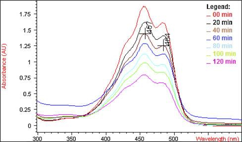
The degradation of BC in hexane by CSS for a period of 120 min was monitored by UV–vis spectrometry. The spectrum of the BC after 20 min clearly showed a reduction of absorbance at 458 and 485 nm when compared with the BC without exposure to CSS, indicating that the BC had undergone degradation (Fig. 1 ).
|
|
|
Fig. 1. UV–vis spectra of β-carotene after exposed to C. sativa smoke. |
After two hours exposure to CSS, the BC was degraded by 22.0%, a mean degradation rate of 0.18% per minute. The degradation of the BC without the presence of CSS (hexane only) at room temperature (25 °C) was 2.0%, that is at an average rate of 0.017% per minute. There was also loss of color in the solution containing BC and hexane, which changed from reddish-orange to orange, indicating a change in the chemical structure of the BC. Based on the degradation kinetic curve, the theoretical calculation indicated that the BC would be completely degraded after 9 h into the experiment (27 smoke injections). In reality, this degradation after 9 h was 98.8%. The corresponding degradation after 9 h of the BC in hexane with injections of air without CSS was 8.4%. The chemical profile obtained by GC–MS of the BC samples after contact with the CSS identified the presence of 13 compounds, representing 81.7% of the sample’s chemical composition. Of these 13 compounds, 6 were related to degradation products of the BC after exposure to CSS (Table 1 ). The major compounds identified firstly as BC degradation products were 3-hydroxy-2,4,4-trimethylpentyl)2-methylpropanoate and (2-ethyl-3-hydroxyhexyl)2-methylpropanoate. In a previous study, the 3-hydroxy-2,4,4-trimethylpentyl)2-methylpropanoate and similar compounds were found at higher levels in the exhalations of colorectal cancer patients [23] . The presence these volatile organic compounds in exhalations from cancer patients is associated with excessive oxidation reactions and high levels of cytochrome P450 activity by lipid peroxidation of polyunsaturated fatty acids [24] . One of the major cytotoxic compounds identified as lipid peroxidation product is the 4-hydroxy-2-nonenal, which is toxic for the liver and the kidneys. The lipid peroxidation causes serious membrane damage and may therefore lead to cell death. Compound antioxidants such as BC and α-tocopherol may avoid peroxidation. Our study, for the first time, clearly showed that BC is degraded by CSS, generating compounds identical to degraded products of lipid content in humans, owing to oxidative stress. Previous studies of BC oxidative cleavage in vitro revealed that the major degradation products identified were β -cyclocitral, β -ionone, ionene, and these β -carotene metabolites products may be associated to the carcinogenic response in the lungs of cigarette smokers [25] . The compound β -cyclocitral product of oxidative cleavage reaction of β -carotene was also identified here. BC degradation products induce genotoxic effects at concentrations which may occur under conditions of intense oxidative stress, e.g. induced by a smoker who smokes an excessive number of cigarettes [26] .
| N° | Compound | Molecular formula | RT min | % | Main fragments (m /z ) |
|---|---|---|---|---|---|
| 1 | (3-Hydroxy−2,4,4-trimethylpentyl)2-methylpropanoatea | C12H24O3 | 19.5 | 30.0 | 43, 56, 71, 83, 98 |
| 2 | (2-ethyl−3-hydroxyhexyl)2-methylpropanoatea | C12H24O3 | 20.5 | 20.1 | 43, 56, 71, 89, 173 |
| 3 | dihydroactinidiolidea | C11H16O2 | 26.2 | 3.0 | 43, 111, 137, 180 |
| β-cyclocitrala | C10H16O | 32.6 | 3.7 | 69, 109, 137, 152 | |
| 4 | Cannabidiol | C21H30O2 | 47.9 | 0.3 | 55, 147, 231, 267 |
| 5 | 2-tridecen−1-ala | C13H24O | 49.2 | 1.0 | 43, 149, 167, 231 |
| 6 | Ergost-5-en-3-ol, acetate, (3β,24R) | C30H50O2 | 49.5 | 0.4 | 43, 55, 231, 367 |
| 7 | Δ9-tetrahydrocannabinol | C21H30O2 | 50.3 | 11.4 | 258, 271, 299, 314 |
| 8 | Cannabigerol | C21H32O2 | 51.2 | 1.0 | 43, 55, 69, 123, 193, 231, 295 |
| 9 | Cannabinol | C21H26O2 | 51.6 | 5.4 | 119, 238, 295, 310 |
| 10 | Hexadecane,6, 11-dipentyl | C26H54 | 53.3 | 1.1 | 43,71, 85, 295, 313 |
| 11 | Farnesol isomer Aa | C15H26O | 53.7 | 3.2 | 55, 69, 81, 95, 295 |
| 12 | Hexatriacontane | C36H74 | 55.1 | 0.9 | 43, 57, 71, 85, 99 |
| 13 | Tetracosane, 9-octyl- | C32H66 | 56.3 | 0.2 | 43, 57, 71, 85 |
| TOTAL | – | – | 81.7 | – |
a. Probable degradation products of BC after exposure to CSS.
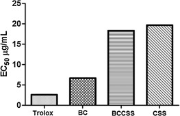
Considering the high degree of degradation of BC by CSS and the known antioxidant activity of BC, possible loss of BC antioxidant potential was investigated. The samples of BC, BCCSS and CSS presented sequestration activity of the radical cation ABTS·+ , with EC50 values varying from 6.67 to 19.70 μg mL−1 (Fig. 2 ). BC (6.67 ± 0.27 μg mL−1) was the substance with the best antiradical activity in relation to the standard, Trolox (2.60 ± 0.27 μg mL−1 ). This result was expected, because the antioxidant property of BC has been widely studied and is well known. Indeed, BC is used as a standard substance in some antioxidant tests. The EC50 result for the BCCSS (18.53 ± 1.1 μg mL−1 ) indicated that the electron sequestration capacity of the BC was reduced by about three times after contact with the CSS. The significant decrease in antioxidant activity of the BC, after contact with the CSS, was due to the BCs chemical structure having undergone chemical changes with a loss of its chemical conjugation and generating non-conjugated degradation products. These compounds with highly conjugated structures, as in case of BC, had high antioxidant potential. The EC50 values of the BCCSS versus CSS (19.70 ± 0.48 μg mL−1 ) presented a small but significant difference between the results, which can confirm the reduction of the antioxidant property of the BC in this sample. This difference is probably related to the small quantity of BC still existing in the composition of the BCCSS sample.
|
|
|
Fig. 2. Graphical plot of effective concentration values (EC50 ) of ABTS radical scavenging activities of the samples of BC, BCCSS and CSS. |
3.2. Assessment of toxicity in the microcrustacean Artemia salina
The purpose of the bioassay with Artemia salina was to obtain information on the toxicity of the samples, including to subsequent toxicological studies. The BC did not cause mortality of A. salina in all the tested concentrations (LC50 > 1000 mg/mL), thus being considered non-toxic. The sample BCCSS presented a LC50 = 397.35 mg/mL, while the sample of CCS presented a LC50 = 461.86 mg/mL. Both samples considered were toxic against A. salina , demonstrating that the BC the compound proved to be toxic after contact with CSS. This is because the CCS degrades the BC, generating oxidative degradation products, such as unsaturated acids, aldehydes, ketones and alcohols, compounds identical to the lipid peroxidation products and generally toxic.
3.3. Assessment of the hemolytic potential in mouse erythrocytes
The samples were tested up to a concentration of 500 μg/mL and none of them caused significant alterations in the erythrocytes’ plasma membranes.
The percentages of hemolyzed cells (%HC) at a concentration of 500 μg/mL were evaluated. None of the samples reached the percentage considered harmful, of 50% (BC = 0.69%; CSS = 5.08%; BCCSS = 16.73%). These results also indicate that the EC50 is above 500 μg/mL. Although the results showed that the samples did not have activity, one comparison deserves mention: the BCCSS had a higher hemolytic percentage than the CSS alone.
3.4. Assessment of the toxicity of repeated doses in rats
We evaluated the behavior of the groups after administration of the daily doses. On the first day, the animals (males and females) that received the CSS and BCCSS presented a reduction in corneal reflex, palpebral ptosis and ambulation, remaining in this state during the first hour. These effects were observed during the first seven days of the experiment after each administration, and might be related to the THC present in the CSS and BCCSS, since this cannabinoid is known for its sedative and somnolence effects. These effects were only observed in the first seven days, probably because of the development of tolerance to the drug [27] , which is a serious problem found in patients treated with cannabinoids. The group of females submitted to BCCSS showed intense irritability and moderate vocalization in the first 20 days of the experiment, before and after administration.
There were no significant differences in water and food consumption and body weight in the treated groups in comparison with the negative control group, demonstrating that the samples probably did not cause any alterations in the animals’ metabolism. With respect to the organ weights of the male rats, no significant alteration was observed in the weights of the heart, liver, kidneys, spleen and stomach (Table 2 ). However, in the females, there was a difference in the liver weight for the BCCSS group. Also, the anatompathological analysis of these organs revealed small whitish spots in the liver of two females treated with BCCSS. These are generally associated with fibrosis, but no changes were observed in the morphology, size or color of these organs. In previous study with rat, whitish markings were found around the terminal hepatic veins on the liver surface after administration of carbon tetrachloride due to white blood cell infiltration [28] . Here, it was not possible to associate the whitish sports found in the liver with a particular type of disease.
| Groups | Animals | Water (ml) | Food (g) | Body (g) | Lung (g) | Heart (g) | Liver (g) | Kidney (g) | Spleen (g) | Stomach (g) |
|---|---|---|---|---|---|---|---|---|---|---|
| Negative control | Male | 181.1±29.9 | 156.4 ± 14.9 | 320.4 ± 3.7 | 1.96 ± 0.23 | 1.23 ± 0.07 | 12.27 ± 0.95 | 2.49 ± 0.14 | 1.02 ± 0.15 | 1.72 ± 0.06 |
| Female | 153.5±30.0 | 99.14 ± 14.0 | 246.9 ± 14.6 | 1.92 ± 0.05 | 1.03 ± 0.07 | 9.82 ± 0.60 | 2.38 ± 0.16 | 0.69 ± 0.07 | 1.75 ± 0.20 | |
| BC | Male | 158.8±17.1 | 144.7 ± 13.4 | 309.7 ± 6.7 | 1.77 ± 0.25 | 1.12 ± 0.06 | 11.06 ± 0.55 | 2.20 ± 0.16 | 0.82 ± 0.08 | 1.68 ± 0.09 |
| Female | 176.9±40.3 | 109.7 ± 24.2 | 258.2 ± 5.1 | 1.92 ± 0.13 | 1.00 ± 0.08 | 9.14 ± 0.79 | 2.34 ± 0.20 | 0.85 ± 0.06 | 1.77 ± 0.19 | |
| CSS | Male | 167.9±30.0 | 141.7 ± 13.8 | 314.2 ± 18.4 | 1.83 ± 0.18 | 1.18 ± 0.15 | 11.82 ± 0.81 | 2.41 ± 0.23 | 0.94 ± 0.04 | 1.81 ± 0.15 |
| Female | 192.5±33.5 | 122.1 ± 25.6 | 251.3 ± 10.8 | 1.82 ± 0.11 | 0.93 ± 0.12 | 9.43 ± 0.70 | 2.14 ± 0.13 | 0.73 ± 0.14 | 1.73 ± 0.10 | |
| BCCSS | Male | 162.9 ± 19.1 | 136.5 ± 18.5 | 318.9 ± 11.9 | 1.88 ± 0.35 | 1.11 ± 0.12 | 11.08 ± 0.56 | 2.37 ± 0.09 | 0.85 ± 0.07 | 1.76 ± 0.07 |
| Female | 197.3 ± 44.5 | 116.6 ± 20.4 | 238.9 ± 5.2 | 1.73 ± 0.24 | 0.91 ± 0.06 | 8.25 ± 0.42 | 2.12 ± 0.27 | 0.68 ± 0.12 | 1.70 ± 0.12 | |
The data represent the mean ± standard deviation. p < 0.05.
BC is carried in the bloodstream by lipoproteins and it is stored mainly in the adipose tissue and liver. Its conversion into retinoids occurs in various organs, such as the liver, lungs and kidneys [29] . Therefore, biochemical analyses of the animals’ blood were chosen referentially to obtain parameters to indicate changes in the organs such as the liver and kidneys. No significant changes were observed in the biochemical parameters analyzed in the male animals (Table 3 ). However, reductions in the concentration of bilirubin and the enzyme alkaline phosphatase were observed in the females that received CSS and BCSS in relation to the control group.
| Biochemical parameters | Animals | Negative control | BC | CSS | BCCSS |
|---|---|---|---|---|---|
| Urea (mg/dL) | Male | 33.45 ± 3.96 | 38.14 ± 4.28 | 38.49 ± 3.24 | 26.70 ± 3.56 |
| Female | 42.80 ± 8.70 | 38.92 ± 9.00 | 22.06 ± 2.2 | 26.10 ± 5.20 | |
| Creatinine (mg/dL) | Male | 0.20 ± 0.07 | 0.28 ± 0.10 | 0.20 ± 0.08 | 0.31 ± 0.09 |
| Female | 0.38 ± 0.10 | 0.32 ± 0.13 | 0.22 ± 0.04 | 0.22 ± 0.10 | |
| AST (U/L) | Male | 79.38 ± 34.71 | 120.3 ± 21.82 | 85.80 ± 20.13 | 76.57 ± 20.13 |
| Female | 67.67 ± 18.5 | 90.22 ± 28.9 | 64.55 ± 23.4 | 62.42 ± 28.60 | |
| ALT (U/L) | Male | 25.57 ± 5.70 | 36.41 ± 4.70 | 31.26 ± 6.9 | 29.39 ± 5.30 |
| Female | 40.14 ± 5.24 | 47.78 ± 14.70 | 27.93 ± 7.64 | 37.92 ± 8.16 | |
| Bilirubin (mg/dL) | Male | 0.05 ± 0.01 | 0.07 ± 0.01 | 0.07 ± 0.01 | 0.07 ± 0.01 |
| Female | 0.19 ± 0.05 | 0.15 ± 0.02 | 0.10 ± 0.03* | 0.08 ± 0.03* | |
| AF (mU/mL) | Male | 132.4 ± 10.21 | 234.0 ± 67.87 | 160.6 ± 26.78 | 207.6 ± 85.80 |
| Female | 316.6 ± 19.02 | 244.9 ± 61.75 | 109.0 ± 14.8* | 211.8 ± 59.50* | |
The data represent the mean ± standard deviation. p < 0.05; *p < 0.01.AST—aspartate aminotransferase; ALT—alanine aminotransferase; AF—alkaline phosphatase.
In adults, carotene supplements such as BC stimulates the immune response by averting low white blood cell counts. However, our data showed that in the analysis of the white blood cells, the groups treated with CSS and BCCSS presented a reduction in the number of leukocytes in the in the females, compared to the control group (Table 4 ).
| Animals | Negative Control | BC | CSS | BCCSS | |
|---|---|---|---|---|---|
| Leukocytes (103/mm3) | Male | 3.60±0.9 | 5.86±2.1 | 8.75±1.1* | 6.76±1.3* |
| Female | 5.08±0.6 | 5.20±1.2 | 3.32±0.6* | 3.37±0.7* | |
| Targeted (%) | Male | 21.46±8.3 | 28.1±3.7 | 24.00±4.2 | 22.58±5.6 |
| Female | 23.25±2.1 | 20.61±4.8 | 19.25±4.1 | 17.62±3.9 | |
| Rods (%) | Male | 1.30±0.7 | 1.60±0.8 | 1.20±0.3 | 2.25±1.3 |
| Female | 0.58±0.5 | 0.53±0.5 | 0.75±0.4 | 0.75±0.4 | |
| Eosinophils (%) | Male | 0.69±0.6 | 0.70±0.5 | 1.10±0.2 | 0.66±0.7 |
| Female | 0.50±0.5 | 0.38±0.4 | 0.75±0.7 | – | |
| Lymphocytes (%) | Male | 68.92±8.2 | 65.7±4.0 | 71.20±4.5 | 79.75±4.7* |
| Female | 70.08±4.1 | 75.92±5.2 | 76.83±5.5 | – | |
| Monocytes (%) | Male | 3.76±1.6 | 3.90±1.1 | 2.70±1.4 | 3.33±1.8 |
| Female | 5.58±3.7 | 2.53±1.3 | 2.41±1.4 | 1.87±0,9 | |
| Basophils (%) | Male | – | – | – | – |
| Female | – | – | – | – | |
The data represent the mean ± standard deviation. p < 0.05; *p < 0.01.
In the assessment of the hematological parameters, no significant differences were observed in the red blood cells, hemoglobin concentration and hematocrit count, and no erythrocyte anomalies were detected (Table 5 ).
| Animals | Negative Control | BC | CSS | BCCSS | |
|---|---|---|---|---|---|
| RCB (106/mm3) | Male | 4.22 ± 1.0 | 4.343 ± 0.8 | 4.742 ± 0.4 | 4.19 ± 1.2 |
| Female | 7.58 ± 0.2 | 7.32 ± 0.3 | 7.70 ± 0.3 | 7.77 ± 0.4 | |
| Hemoglobin (g/dL) | Male | 7.68 ± 1.7 | 8.050 ± 1.5 | 8.860 ± 0.8 | 8.58 ± 0.8 |
| Female | 14.27 ± 0.3 | 13.93 ± 0.5 | 14.70 ± 0.7 | 14.16 ± 0.8 | |
| Hematocrit (%) | Male | 21.63 ± 5.4 | 22.78 ± 4.4 | 24.46 ± 2.3 | 23.86 ± 2.5 |
| Female | 42.45 ± 1.3 | 41.50 ± 1.5 | 43.16 ± 2.1 | 42.00 ± 2.2 | |
| MCV (m3) | Male | 51.14 ± 1.5 | 52.33 ± 1.9 | 51.40 ± 0.9 | 51.00 ± 1.5 |
| Female | 56.00 ± 1.5 | 56.67 ± 2.0 | 56.14 ± 1.3 | 53.90 ± 1.1 | |
| MCH (g.10−12) | Male | 18.21 ± 0.9 | 18.52 ± 0.4 | 18.64 ± 0.2 | 18.52 ± 0.2 |
| Female | 18.72 ± 0.4 | 19.00 ± 0.7 | 19.10 ± 0.6 | 18.20 ± 0.4 | |
| MCHC (g/dL) | Male | 35.70 ± 1.6 | 35.42 ± 0.7 | 36.16 ± 0.2 | 36.25 ± 0.9 |
| Female | 33.67 ± 0.8 | 33.58 ± 0.1 | 34.06 ± 0.8 | 33.72 ± 0.6 | |
| RDW (%) | Male | 13.59 ± 0.6 | 14.37 ± 1.2 | 13.34 ± 0.3 | 13.57 ± 0.7 |
| Female | 14.78 ± 0.4 | 14.23 ± 0.8 | 14.60 ± 0.5 | 14.02 ± 0.7 | |
The data represent the mean ± standard deviation p < 0.05. RBC—red blood cells; MCV— mean corpuscular volume; MCH—mean corpuscular hemoglobin; MCHC—mean corpuscular hemoglobin concentration; RDW—red cell distribution width.
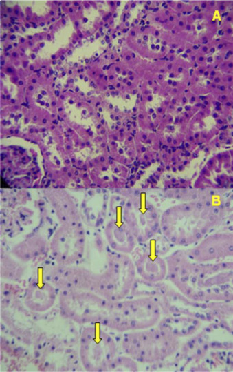
In the histopathological analysis of the organs, only the kidneys of the male and female rats treated with BCCSS presented alterations. In the kidneys of these animals, the cortical region contained amorphous eosinophilic material (cylinders) in the lumen of the proximal and distal convoluted tubules (Fig. 3 ). Discrete lymphoplasmacytic interstitial nephritis was also observed in the groups treated with CSS and BCCSS.
|
|
|
Fig. 3. Cortical region of the kidneys of male Wistar rats submitted to 30 days of exposure A—kidney of the control group; B—kidney of the BCCSS group (male rat); C—kidney of the BCCSS group (female rat). *yellow arrow—amorphous eosinophilic material. |
The presence of cylinders can be related to various factors, such as problems of protein reabsorption, nephritis and renal tubular lesion. This can indicate toxicity, since the formation of intratubular cylinders can obstruct the tubules and impair their function [30] . However, other examinations need to be conducted to establish the origin of these cylinders.
4. Conclusion
The BC is an important natural carotenoid for human health by acting to prevent various diseases including lung cancer and as it is a precursor of vitamin A. Its chemical structure with forty carbon atoms and the fact that it is highly conjugated makes it sensitive to a degradation that can generate various potentially toxic compounds. In this study we show that the CSS exposed to BCC undergoes changes in its chemical structure, altering its biological properties and losing its antioxidant potential by about three times. In rats, although females were more sensitive, in both sexes, after repeated doses of BCCSS, the kidneys showed discrete amorphous eosinophilic material (cylinders) in the lumen of the proximate and distal convoluted tubules, besides discrete lymphoplasmacytic interstitial nephritis. Thus, this study shows that BC intake together with CSS should not be encouraged due to toxicity and loss of antioxidant activity of the BC after contact with the CSS.
Conflict of interest
The authors have no conflict of interest to report.
Transparency document
Acknowledgments
The authors are indebted to the Centro de Apoio a Pesquisa (CENAPESQ), UFRPE, and to the Pernambuco Police Science Laboratory, Brazil, for the laboratory facilities. Authors thanks also to the Judge José Claudionor da Silva Filho and Dr. Luiz Carlos Soares da Silva for authorization and providing of C. sativa .
Appendix A. Supplementary data
The following are Supplementary data to this article:
References
- [1] J. Fiedor, K. Burda; Potential role of carotenoids as antioxidants in human health and disease; Nutrients, 6 (2014), pp. 466–488
- [2] J.A. Olson; Absorption, transport, and metabolism of carotenoids in humans; Pure Appl. Chem., 66 (1994), pp. 1011–1016
- [3] G.R. Chichili, D. Nohr, M. Saffer, J. Lintig, H.K. Biesalski; Carotene conversion into vitamin a in human retinal pigment epithelial cells; IOVS, 46 (2005), pp. 3562–3569
- [4] K. Briviba, K. Schnäbele, E. Schwertle, M. Blockhaus, G. Rechkemmer; β-Carotene inhibits growth of human colon carcinoma cells in vitro by induction of apoptosis; Biol. Chem., 382 (2005), pp. 1663–1668
- [5] T. Sacha, M. Zawada, J. Hartwich, Z. Lach, A. Polus, M. Szostek, E. Zdzi Owska, M. Libura, M. Bodzioch, A. Dembińska-Kieć, A.B. Skotnicki, R. Góralczyk, K. Wertz, G. Riss, C. Moele, T. Langmann, G. Schmitz; The effect of β -carotene and its derivatives on cytotoxicity, differentiation, proliferative potential and apoptosis on the three human acute eukemia cell lines: u-937 HL-60 and TF-1 ; Biochim. Biophys. Acta, 1740 (2005), pp. 206–214
- [6] Y. Cui, Z. Lu, L. Bai, Z. Shi, W.E. Zhao, B. Zhao; β -Carotene induces apoptosis and up-regulates peroxisome proliferator-activated receptor γ expression and reactive oxygen species production in MCF-7 cancer cell ; Eur. J. Cancer, 43 (2007), pp. 2590–2601
- [7] G.S. Omenn; Risk factors for lung cancer and for intervention effects in CARET, the beta-carotene and retinol efficacy trial; J. Natl. Cancer Inst., 334 (1996), pp. 1550–1559
- [8] A. Rahman, S.S.I. Bokhari, M.A. Waqar; Beta-carotene degradation by cigarette smoke in hexane solution in vitro; Nutr. Res., 21 (2001), pp. 821–829
- [9] P. Palozza, S. Serini, S. Trombino, L. Lauriola, F.O. Ranelletti, G. Calviello; Dual role of β -carotene in combination with cigarette smoke aqueous extract on the formation of mutagenic lipid peroxidation products in lung membranes: dependence on pO2 ; Carcinogenesis, 27 (2006), pp. 2383–2391
- [10] G.M. Lowe, K. Vlismas, D.L. Graham, M. Carail, C. Caris-Veyrat, A.J. Young; The degradation of (all-E ) β -carotene by cigarette smoke ; Free Radic. Res., 43 (2009), pp. 280–286
- [11] M. Hashibe, K. Straif, D.P. Tashkin, H. Morgenstern, S. Greenland, Z.F. Zhang; Epidemiologic review of marijuana use and cancer risk; Alcohol, 35 (2005), pp. 265–275
- [12] K.M. Honório, A. Arroio, A.B.F. Silva; Aspectos terapêuticos de compostos da planta Cannabis sativa; Quím. Nova, 29 (2006), pp. 318–325
- [13] Z. Bruci; First systematic evaluation of the potency of Cannabis sativa plants grown in Albania ; Forensic Sci. Int., 10 (2012), pp. 40–46
- [14] W. Hall; The mental health risks of adolescent Cannabi s use ; PLoS Med., 3 (2006), p. e39
- [15] A.J. Gordon, J.W. Conley, J.M. Gordon; Medical consequences of marijuana use: a review of current literature; Curr. Psychiatry Rep., 15 (2013) (419-11)
- [16] L. Degenhardt, A. Roxburgh, R. Mcketin; Hospital separations for cannabis and methamphetamine-related psychotic episodes in Austrália; Med. J. Aust., 186 (2007), pp. 342–345
- [17] H. Friedman, S. Pross, T.W. Klein; Addictive drugs and their relationship with infectious diseases FEMS Immunol; Med. Microbiol., 47 (2006), pp. 330–342
- [18] R. Re, N. Pellegrini, A. Proteggente, A. Pannala, M. Yang, C. Rice-Evans; Antioxidant activity applying an improved ABTS radical cation decolorization assay; Free Radic. Biol. Med., 26 (1999), pp. 1231–1237
- [19] L.V. Costa-Lotufo, G.M. Cunha, P.A. Farias, G.S. Viana, K.M. Cunha, C. Pessoa, M.O. Moraes, E.R. Silveira, N.V. Gramosa, V.S. Rao; The cytotoxic and embryotoxic effects of kaurenoic acid, a diterpene isolated from Copaifera langsdorffii oleo-resin; Toxicon, 40 (2002), pp. 1231–1234
- [20] J.S. Aguiar, R.O. Araújo, M.D. Rodrigues, K.X.F.R. Sena, A.M. Batista, M.M.P. Guerra, S.L. Oliveira, J.F. Tavares, M.S. Silva, S.C. Nascimento, T.G. da Silva; Antimicrobial, antiproliferative and proapoptotic activities of extract fractions and isolated compounds from the stem of erythroxylum caatingae plowman; Int. J. Mol. Sci., 13 (2012), pp. 4124–4140
- [21] R.N. Almeida, A.C.G.M. Falcão, R.S.T. Diniz, L.J. Quintans-Júnior, R.M. Polari, J.M. Barbosa-Filho, M.F. Agra, J.C. Duarte, C.D. Ferreira, A.R. Antoniolli, C.C. Araújo; Metodologia para avaliação de plantas com atividade no sistema nervoso central e alguns dados experimentais; Ver. Bras. Farm., 80 (1999), pp. 72–76
- [22] B.N. Meyer, N.R. Ferrigni, J.E. Putnam, L.B. Jacobsen, D.E. Nichols, J.L. McLaughlin; Brine shrimp: a convenient general bioassay for active plant constituents; Planta Med., 45 (1982), pp. 31–34
- [23] C. Wang, C. Ke, X. Wang, C. Chi, L. Guo, S. Luo, Z. Guo, G. Xu, F. Zhang, E. Li; Noninvasive detection of colorectal cancer by analysis of exhaled breath; Anal. Bioanal. Chem., 406 (2014), pp. 4757–4763
- [24] M. Phillips, R.N. Cataneo, C. Saunders, P. Hope, P. Schmitt, J. Wail; Volatile biomarkers in the breath of women with breast cancer; J. Breath Res., 4 (2010), p. 026003 (8pp)
- [25] O. Sommerburg, N. Karius, W. Siems, C. Langhans, M. Leichsenring, N. Breusing, T. Grune; Proteasomal degradation of β -carotene metabolite-modified proteins ; BioFactors, 35 (2009), pp. 449–459
- [26] A.J. Alija, N. Bresgen, O. Sommerburg, W. Siems, P.M. Eckl; Cytotoxic and genotoxic effects of β -carotene breakdown products on primary rat hepatocytes ; Carcinogenesis, 25 (2004), pp. 827–831
- [27] V.M. Saito, C.T. Wotjak, F.A. Moreira; Exploração farmacológica do sistema endocanabinoide: novas perspectivas para o tratamento de transtornos de ansiedade e depressão?; Ver Bras. Psiquiatr., 32 (2010), pp. 7–14
- [28] T. Ito, T. Itoshima, S. Kiyotoshi, K. Kawaguchi, H. Ogawa, S. Hattori, M. Kitadai, T. Maruyama, H. Nagashima; Peritoneoscopic study on rat liver cell necrosis induced by carbon tetrachloride and allyl formate; Gastroenterol. Jpn., 17 (1) (1982), pp. 31–35
- [29] T. Grune, G. Lietz, A. Palou, A.C. Ross, W. Stahl, G. Tang, D. Thurnham, S. Yin, H.K. Biesalski; β -Carotene is an important vitamin A source for humans ; J. Nutr, 22 (2010), pp. 2268S–2285S
- [30] G. Camusi, C. Ronco, G. Montrucchio, G. Piccoli; Role of soluble mediators in sepsis and renal failure; Kidney Int. Suppl., 54 (1998), pp. 38–42
Document information
Published on 02/05/17
Accepted on 02/05/17
Submitted on 02/05/17
Licence: Other
Share this document
Keywords
claim authorship
Are you one of the authors of this document?


