Summary
Cancer of the appendix is very rare and is typically found incidentally in approximately 1% of patients who received appendectomies. Most of appendiceal tumors are carcinoid, adenoma, and lymphoma. Adenocarcinoma of the appendix accounts for only 10% of all primary appendiceal cancers and the treatment remains controversial. In this article, we report a 67-year-old man who presented with symptoms of appendicitis that was diagnosed as adenocarcinoma of the appendix Stage II. The patient was treated with right hemicolectomy. To date he remains asymptomatic.
Keywords
Appendix adenocarcinoma; Appendix tumor; Appendicitis
Introduction
The appendix is thought to play a role in the immune system. However, the real function of the appendix in humans is unclear. The appendix may become cancerous in some rare conditions. The majority of appendiceal cancers are carcinoids, while the remaining 10–20% are mucinous cystadenocarcinoma, adenocarcinoma, lymphosarcoma, paraganglioma, and granular-cell tumors [1]. Most appendiceal cancers present symptoms of appendicitis, including fever, leukocytosis, and acute abdominal pain in the right lower quadrant. Sometimes, patients may present chronic abdominal pain just like the case we are reporting. Appendectomy is performed as clinically indicated. Sometimes, an appendiceal mass is found incidentally during abdominal surgery. Most masses are benign mucoceles or very small carcinoids and they do not require any further management [1]. Rarely, when the mass is diagnosed as adenocarcinoma, the treatment algorithm remains controversial [1].
Case Report
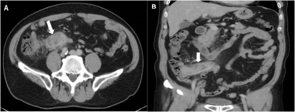
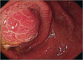
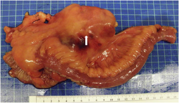
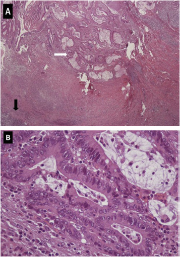
A 67-year-old man came to the outpatient department with intermittent right lower abdominal pain ongoing for 1 month. He denied any systemic disease and had no fever, chills, hematochezia, chronic diarrhea, nor body weight loss. Physical examination revealed right lower quadrant tenderness without rebound pain and muscle guarding. Positive psoas sign was noted. Blood tests were unremarkable except mild elevated C-reactive protein (4.9 mg/dL). Abdominal computed tomography (CT) demonstrated marked appendiceal wall swelling and intraluminal filling defect of contrast medium over the appendix (Figure 1). The colonoscopy revealed a protruding mass with edematous mucosa at cecum (Figure 2). Biopsy obtained at colonoscopy revealed inflammation of appendix mucosa. Due to the cancer could not be ruled out, a surgery with right hemicolectomy was performed and which showed a 7-cm length appendiceal tumor (Figure 3). Hematoxylin and eosin staining showed irregular glands with stromal invasion to muscularis propria, intraluminal mucin extravasation, and abscess formation were also seen (Figure 4A). Glandular arrangement lining by pseudostratified columnar cells with enlarged vesicular nuclei was noted under high magnification (Figure 4B). On the basis of these findings, he was diagnosed as colonic type adenocarcinoma of the appendix, T2N0M0, Stage I. After right hemicolectomy, he remains asymptomatic to date.
|
|
|
Figure 1. Computed tomography images: (A) cross section of abdominal computed tomography showed an appendiceal tumor (white arrow); and (B) marked appendiceal wall swelling and intraluminal filling defect of contrast medium over appendix noted (white arrow). |
|
|
|
Figure 2. A protruding mass with edematous mucosa noted at cecum. |
|
|
|
Figure 3. A surgery with right hemicolectomy showed a 7-cm length appendiceal tumor (white arrow). |
|
|
|
Figure 4. (A) Irregular glands with stromal invasion to muscularis propria noted under H&E stain (×40 original magnification); intraluminal mucin extravasation (white arrow) and abscess formation (black arrow) were also seen. (B) High magnification under H&E stain showed glandular arrangement lining by pseudostratified columnar cells with enlarged vesicular nuclei (×100 original magnification). Colonic type adenocarcinoma of appendix was diagnosed. |
Discussion
Adenocarcinoma of the appendix is a rare disease, which accounts for 0.5% of all gastrointestinal cancers [2]. The peak incidence of appendix adenocarcinoma is in the 6th decade with a slight male predominance [3]. Most patients with appendiceal adenocarcinoma present with acute appendicitis, probably due to early malignant obstruction of the appendiceal lumen leading to superimposed infection [3].
CT imaging is helpful in identifying a possible appendiceal neoplasm. Calcifications may indicate this process is malignant rather than infective or inflammatory. There may be some air bubbles noted on the CT image if there is a superinfection associated with the malignancy. In certain conditions, such as abdominal distension caused by pseudomyxoma peritonei, a CT would show widespread heterogeneous locules in the peritoneal cavity [4]. Since there are no imaging studies specific for diagnosing adenocarcinomas, the final diagnosis is most often made postoperatively on microscopic examination.
Histological classification is into either mucinous (55%), colonic (34%), or adenocarcinoid types (11%) [1]. Once diagnosed, it has been generally accepted that a right hemicolectomy is the preferred surgical intervention with a clear survival benefit [1] and [3]. While there are small studies and anecdotal case reports that suggest a response to regimens containing fluorouracil, the role of chemotherapy has yet to be clearly defined. Generally, systemic and intraperitoneal chemotherapy should be used in the presence of nodal involvement [2], [5] and [6].
Prognosis of adenocarcinoma depends on the subtype and extent of disease. Those patients would have a worse prognosis when intraperitoneal seeding occurred. Mucinous adenocarcinoma has a more favorable prognosis because it does not exhibit hematogenous or lymphatic spread [5]. Goblet cell subtype has a worse prognosis with one study listing a 55% 5-year survival rate [6]. Some studies have shown that there is a clear survival benefit to the addition of a hemicolectomy. In a study by Nilecki et al [2], the 5-year survival rate for hemicolectomy was 73% versus 44% in the appendectomy group.
In conclusion, adenocarcinoma of the appendix is rare and it may present as appendicitis. Right hemicolectomy should be performed once diagnosed.
Conflicts of interest
We faithfully and honestly disclose that this work has not been presented in any form or published by any journal. There is no conflict of interest for any of the authors.
References
- [1] C. Ruoff, L. Hanna, W. Zhi, G. Shahzad, V. Gotlieb, M. Wasif Saif; Cancers of the appendix: review of the literatures; ISRN Oncol, 2011 (2011), p. 728579
- [2] S.S. Nilecki, B.G. Wolff, R. Schlinkert, M.G. Sarr; The natural history of surgically treated primary adenocarcinoma of the appendix; Ann Surg, 219 (1994), pp. 51–57
- [3] J.E. Hartley, P.J. Drew, A. Qureshi, A. MacDonald, J.R.T. Monson; Primary adenocarcinoma of the appendix; J Roy Soc Med, 89 (1996), pp. 111–113
- [4] P.J. Pickhardt, A.D. Levy, C.A. Rohrmann Jr., A.I. Kende; Primary neoplasms of the appendix: radiologic spectrum of disease with pathologic correlation; Radiographics, 23 (2003), pp. 645–662
- [5] P.S. Pahlavan, R. Kanthan; Goblet cell carcinoid of the appendix; World J Surg Oncol, 3 (2005), p. 36
- [6] P.H. Sugarbaker, K. Kern, E. Lack; Malignant pseudomyxoma peritonei of colonic origin. Natural history and presentation of a curative approach to treatment; Dis Colon Rectum, 30 (1987), pp. 772–779
Document information
Published on 31/08/16
Licence: Other
Share this document
Keywords
claim authorship
Are you one of the authors of this document?


