(Created page with "==Summary== Transposition of the gallbladder to the left side without ''situs inversus viscerum'' is rare. These gallbladders are situated under the left lobe of the liver be...") |
m (Scipediacontent moved page Draft Content 897076759 to Makni et al 2012a) |
(No difference)
| |
Latest revision as of 11:11, 26 May 2017
Summary
Transposition of the gallbladder to the left side without situs inversus viscerum is rare. These gallbladders are situated under the left lobe of the liver between Segment III and IV or on Segment III to the left of the falciform ligament. This is a report of a 50-year-old woman who was admitted to our department with a history of pain in her right upper abdomen. The physical examination showed tenderness in the right upper quadrant of the abdomen without a Murphys sign. Abdominal ultrasonography showed gall bladder stones without dilatation of the bile ducts. The patient underwent a laparoscopic cholecystectomy using the French position and four ports positioned as usual. We discovered a left-sided gallbladder located on the left of the round ligament. The gallbladder was excised as usual. Intraoperative cholangiogram showed neither dilatation of the bile ducts nor associated congenital anomalies of the biliary tree. The patient was discharged on the first postoperative day. Because routine preoperative examinations may not detect the anomaly, the latter may take surgeons by surprise during laparoscopy. Awareness of the unpredictable confluence of the cystic duct into the common bile duct and selective use of intraoperative cholangiography both contributed to the safe laparoscopic management of this unusual problem.
Keywords
aberrant gallbladder;laparoscopy;left-sided gallbladder
1. Introduction
Patients with congenital anomalies of the biliary tree are often met in daily clinical practice. Left-sided gallbladder is an uncommon anatomic abnormality defined as a gallbladder located on the left side of the round ligament. We report herein a case of left-sided gallbladder discovered incidentally during laparoscopic cholecystectomy.
2. Case report
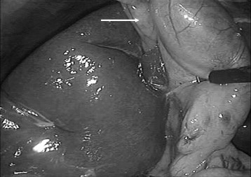
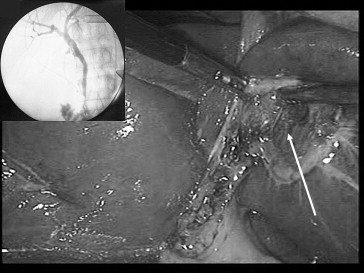
A 50-year-old woman with no medical history was admitted to our department with a history of 2 years' intermittent pain in her right upper abdomen, radiating to the back. There was no history of jaundice. The physical examination showed tenderness in the right upper quadrant of the abdomen without a Murphys sign. Laboratory findings demontrated normal rates of total bilirubin, alkaline phosphatase, γ-glutamyl transpeptidase, and C-reactive protein. Abdominal ultrasonography showed gallbladder stones without dilatation of the bile ducts. The patient underwent a laparoscopic cholecystectomy using the French position and four ports positioned as usual. The gallbladder was absent from its normal location. Medial exploration of the falciform ligament under the left liver revealed the gallbladder attached to a shallow fossa in the liver (Fig. 1). The cystic duct joined the common hepatic duct on the right side after making a hairpin bend, and the cystic artery originated from the right hepatic artery. Retrograde cholecystectomy was undertaken using electrocautery followed by intraoperative cholangiogram which showed neither dilatation of the bile ducts nor associated congenital anomalies of the biliary tree (Fig. 2). The patient was discharged on the first postoperative day, and had an uneventful postoperative period. Histological examination showed chronic cholecystitis.
|
|
|
Figure 1. Gallbladder lying to the left of the falciform ligament (white arrow) in relation to Segment III. |
|
|
|
Figure 2. After cholecystectomy, the photo shows the gallbladder bed (White arrow) to gauci of falciform ligament. Inset: The intraoperative cholangiography shows a biliary modal mapping. |
3. Discussion
The reported prevalence of left-sided gallbladder is 0.1% to 0.7%.1 It was first described by Hochstetter2 in 1886. A review of the literature shows that to date, about 149 cases of left-sided gallbladder have been reported,3 the most recent being published by Kelly et al in 2008.4
Goss et al have classified aberrant gallbladders in four sites: intrahepatic, left-sided, retrodisplaced, and transverse.5 The exact etiology of this anomaly is uncertain. It may be caused by the gallbladder bud arising from the hepatic diverticulum and migrating to the left lobe instead of the right. This migration explains the entry of the cystic duct on the right side of the hepatic duct, as described in our case.5 ; 6 Another explanation of this variation may be the development of a second gallbladder directly from the left hepatic duct, accompanied by a failure of absence of the normal gallbladder on the right side.5 Some left-sided gallbladders have been reported by Nagai et al who found the anomaly associated with a right-sided falciform ligament in 3 out of 1621 patients (0.2%) being operated.7 This anomaly should be suspected when ultrasonography, computed tomography, and angiography reveal no definite Segment IV, absence of umbilical portion of the portal vein in the left lobe, and club-shaped right anterior portal vein.8 However,preoperative ultrasonography may not detect the anomaly, as was the case for our patient. We concluded that we should have performed an additional intraoperative ultrasonography to detect these portal vein anomalies. There are several therapeutic implications of an aberrant gallbladder beneath the left lobe of liver. Left-sided gallbladder may be associated with the anomalies of the intrahepatic portal vein (trifurcation type),8 ; 9 or the cystic duct,9 or with an accessory liver10 the detection of this congenital anomaly of the biliary has several therapeutic implications especially in cases of hepatic resection.
Laparoscopic intervention is safe when performing cholecystectomy in such a situation.11; 12; 13 ; 14 The first laparoscopic cholecystectomy for left-sided gallbladder was reported by Schiffino et al in 1993 who advised, in this case, performing the cholecystectomy by the anterograde approach in order to visualize the anatomic structures better and safely avoid injury of hepatic pedicle.11 However, in case of difficulties, open surgery should be considered. Adopting the French position, which was the way we choose for our patient, may facilitate the procedure. Some authors have suggested several modifications of the laparoscopic procedure, especially in the ports position and the standing position of operating surgeon. Intraoperative cholangiography should be performed in order to detect other associated anomalies and to define the biliary tree or to diagnose an intraoperative common bile duct injury.
4. Conclusion
Left-sided gallbladder is a rare operative finding. Preoperative imagery may not detect the anomaly. Laparoscopic intervention is safe in such situation. Intraoperative cholangigraphy should be performed to detect associated anomalies of the biliary tree. When the surgeon is doubtful, conversion is recommend to avoid complications.
References
- 1 R. Si-Youn, J. Poong-Man; Left-sided gallbladder with right-sided ligamentum teres hepatis: rare associated anomaly of exomphalos; J Pediatr Surg, 43 (2008), pp. 1390–1395
- 2 F. Hochstetter; Anomaliem der pfortader und der nabelvene in verbindung mit defect oder Linkscage der gall-en-blase; Arch Anat Entwick (1886), pp. 369–384
- 3 A. Dhulkotia, S. Kumar, V. Kabra, H.S. Shukla; Aberrant gallbladder situated beneath the left lobe of liver; HPB, 4 (1) (2002), pp. 39–42
- 4 M.D. Kelly, J.D. Craik; Left-sided gall bladder revisited; ANZ J Surg, 78 (2008 Mar), pp. 192–193
- 5 R.E. Gross; Congenital anomalies of the gallbladder; Arch Surg, 32 (1936), pp. 131–162
- 6 E.A. Boyden; The accessory gallbladder. An embryological and comparative study of aberrant biliary vesicles occurring in a man and the domestic mammals; Amer J Anat, 38 (1926), p. 177
- 7 M. Nagai, K. Kubota, S. Kawasaki, T. Takayama, Y. Bandai, M. Makuuchi; Are left-sided gallbladders really located on the left side?; Ann Surg, 225 (1997), pp. 274–280
- 8 S.L. Hsu, T.Y. Chen, T.L. Huang, et al.; Left-sided gallbladder: its clinical significance and imaging presentations; World J Gastroenterol, 13 (2007), pp. 6404–6409
- 9 A. Ikoma, K. Tamaka, N. Hamada; Left sided gallbladder with accessory liver accompanied by intra-hepatic cholangiocarcinoma; J Jpn Surg Soc, 93 (1992), pp. 434–436
- 10 T. Ogawa, S. Ohwada, S. Ikeya, H. Shiozaki, S. Aiba, Y. Morishita; Left sided gallbladder with anomalies of the intrhepatic portal vein and anomalous junction of the pancreatico-biliary ductal system: a case report; Hepatogastroenterol, 42 (1995), pp. 645–649
- 11 L. Schiffino, J. Mouro, H. Levard, F. Dubois; A case of a left-sided gallbladder treated surgically via laparoscopy; Ann Ital Chir, 64 (1993), pp. 229–231
- 12 N. Hopper, J.M. Ryder, K. Swarnkar, B.M. Stephenson; Laparoscopic left hepatic lobe cholecystectomy; J Laparoendosc Adv Surg Tech A, 13 (2003), pp. 405–406
- 13 N. Matsumura, H. Tokumura, A. Yasumoto, et al.; Laparoscopic cholecystectomy and common bile duct exploration for cholecystocholedocholithiasis with a left-sided gallbladder: report of a case; Surg Today, 39 (2009), pp. 252–255
- 14 N. Kanazumi, M. Fujiwara, H. Sugimoto, et al.; Laparoscopic cholecystectomy for left-sided gallbladder: report of two cases; Hepatogastroenterology, 54 (2007), pp. 674–676
Document information
Published on 26/05/17
Submitted on 26/05/17
Licence: Other
Share this document
Keywords
claim authorship
Are you one of the authors of this document?

