Keywords
Cardiac injury;Thoracentesis catheter
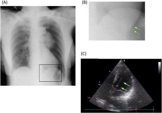
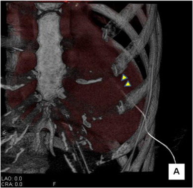
A 73-year-old woman received a thoracoscopic fundoplication for a hiatal hernia and her pleura was partially broken. A chest X-ray showed a pneumothorax in her left lung (Fig. 1(A)). A 6 French aspiration catheter (Trocar Aspiration Catheter Kit®, Nihon Covidien Co, Ltd., Shizuoka, Japan) was inserted into her thorax, resulting in aspirated pulsatile arterial blood. The catheter was quickly clamped after the incorrect insertion. She was transferred to our hospital under intubation for investigations and treatment. Her blood pressure was 122/80 mmHg, and her heart rate was 68 beats per minute without cardiovascular agents. A non-contrast prospective electrocardiogram-triggered computed tomography (CT) scanning (Fig. 2) and echocardiogram (Fig. 1(C)) revealed that the catheter had penetrated the anterior wall in the apex of her myocardium, and the tip was present in the basal portion of her left ventricle (LV) cavity. Fortunately, the side holes in the catheter were all present in the LV cavity (Fig. 2, arrowhead), and there was no evidence of a hematoma in the fluid storage in her pericardium or hemothorax. She was diagnosed with a cardiac trauma and an emergency operation was performed.
|
|
|
Fig. 1. (A) (B)Chest X-ray in former hospital. The catheter seemed to be penetrated her heart (arrow). (C)Cardiac perforation revealed by echocardiogram. The catheter has penetrated the anterior wall in the apex of her myocardium and the tip is present in the basal portion of her left ventricle cavity (arrow). |
|
|
|
Fig. 2. Cardiac perforation revealed by non-contract 3-dimension computed tomography. The catheter has penetrated the anterior wall in the apex of her myocardium and the tip is present in the basal portion of her left ventricle cavity. The side holes in the catheter were all present in her left ventricle cavity (arrowhead). |
After a median sternotomy and opening of the pericardium, the foreign object penetrating the left ventricle was removed. Upon removal, there was a spot in the myocardium near the diagonal branches that continued to bleed and a pin-hole puncture point inside. Finally, the defect in the wall was repaired.
The main features of the present study are as follows: 1) a thoracentesis catheter can perforate the left ventricle; 2) in this case, the patient was transferred to our hospital without removing the thoracentesis catheter. To the best of our knowledge, this is the first report that describes a case of cardiac perforation by a thoracentesis catheter.
The American Association for Surgery Trauma Cardiac Organ Injury Scale (AAST-OIS) divides traumatic cardiac injuries into 5 groups according to severity. The mortality of heart injuries have previously been reported depending on the AAST-OIS [1]. The mortality of heart injuries of an AAST-OIS grade II (penetrating tangential myocardial wound that does not extend through the endocardium, without tamponade) was calculated to be 50% [2].
The most important prognostic factors in cardiac trauma situations are the vital signs of the patient upon arrival at the hospital [2] ; [3]. Furthermore, the cause of trauma (e.g., stab wound or gunshot wound) and presence of cardiac tamponade is also important prognosticators [2] ; [3].
Echocardiograms have been reported to be effective in diagnosing patients with a penetrating cardiac injury, and therapeutic outcomes have been shown to be improved by early diagnosis [4]. In this case, her hemodynamics were stable, and the chest X-rays from the previous hospital demonstrated LV penetration, an echocardiogram was performed to evaluate cardiac functions. However, in emergency cases, echocardiograms would be the first choice for diagnosis.
The possible factors were speculated as follows: in this case, the incorrect insertion was derived from an inadequate visual field due to surgical operation wounds and a hasty procedure to attempt to recover an iatrogenic pneumothorax. Although the physician who performed the thoracentesis is experienced and had previously performed many successful thoracentesis procedures, it is necessary to pay attention in such adverse conditions.
Penetrating cardiac traumas are comparatively rare. Even if hemodynamics are stable, it is easy to fall into cardiac tamponade. Even though rapid diagnosis and treatments are required, it is important to be careful. The patient provided consent for publication.
Funding sources
None.
Potential conflict of interest
None.
References
- [1] E.E. Moore, M.A. Malangoni, T.H. Cogbill, S.R. Shackford, H.R. Champion, G.J. Jurkovich, et al.; Organ injury scaling IV: thoracic vascular, lung, cardiac, and diaphragm; J. Trauma Acute Care Surg., 36 (1994), pp. 299–300
- [2] J.A. Asensio, J.D. Berne, D. Demetriades, L. Chan, J. Murray, A. Falabella, et al.; One hundred five penetrating cardiac injuries: a 2-year prospective evaluation; J Trauma, 44 (1998), pp. 1073–1082
- [3] R.F. Buckman Jr., M.M. Badellino, L.H. Mauro, J.A. Asensio, C. Caputo, J. Gass, et al.; Penetrating cardiac wounds: prospective study of factors influencing initial resuscitation; J Trauma, 34 (1993), pp. 717–725 (discussion 25–7)
- [4] A.N. Patel, C. Brennig, J. Cotner, M.A. Lovitt, M.L. Foreman, R.E. Wood, et al.; Successful diagnosis of penetrating cardiac injury using surgeon-performed sonography; Ann. Thorac. Surg., 76 (2003), pp. 2043–2046 (discussion 6–7)
Document information
Published on 19/05/17
Submitted on 19/05/17
Licence: Other
Share this document
Keywords
claim authorship
Are you one of the authors of this document?

