Abstract
Glucose-6-phosphate dehydrogenase (G6PD), a key regulatory enzyme of the pentose phosphate pathway, catalyses the first rate-limiting reaction to produce ribose-5-phosphate for nucleic acid synthesis and NADPH to use in reductive biosynthesis. The available studies indicate an antioxidant role for G6PD and variation in its levels as a result of cellular insult. In this study, the activity of G6PD was monitored during Nʹ-nitrosodiethylamine (NDEA)-induced hepatic damage in Wistar rats. NDEA generates hepatotoxicity and possesses mutagenic effects. To induce hepatic damage, NDEA was administered at doses of 100 mg kg− 1 body weight week− 1 (i.p.) for 14 days. The animals of the control and treated groups were sacrificed each week. The extent of liver damage was ensured by LFT biomarkers, such as transaminases, ALP, bilirubin and the hepato-somatic index (HSI) along with histopathological observations of H&E and Massons trichrome stained liver specimens. The results of the present study show that at the selected doses, NDEA significantly elevates LFT proteins and bilirubin and damages the lobular architecture in a dose-dependent manner. Software analysis of zymograms indicates maximum activity of the hepatic G6PD levels in day-14 NDEA-treated animals. Our spectrophotometry data further support the above findings on hepatic G6PD levels and demonstrate an approximately 1.63 × and 1.66 × fold increase in day-7 and day-14 NDEA intoxicated animals (P < 0.05). It is concluded that elevation in the G6PD activity is apparently the consequence of NDEA-induced intoxication or oxidative stress, leading to hepatic damage to provide sufficient NADPH for microsomal detoxification and ribose-5-phosphate for DNA synthesis and repair, respectively, to maintain the cellular redox status.
Keywords
Antioxidant ; Glucose-6-phosphate dehydrogenase (G6PD) ; Hepatic injury ; Liver function ; Nʹ-nitrosodiethylamine (NDEA)
Introduction
Glucose-6-phosphate dehydrogenase (G6PD), also known as β-d -glucose-6-phosphate, is a housekeeping enzyme (EC 1.1.1.49) of the pentose phosphate pathway (PPP) and is highly important in a variety of cellular processes (Ibrahim et al., 2014 ). In humans, the monomer of G6PD contains 514 amino acids with a molecular weight of 59 kDa (Tian et al., 1998 ). This key regulatory enzyme, G6PD, catalyses the first rate limiting reaction in the PPP, consequently producing ribose-5-phosphate for nucleic acid synthesis and NADPH for reductive biosynthesis (Shannon et al., 2000 ). Primarily, there are two types of regulation of the PPP: First in the liver, concentrations of both G6PD and 6-phosphogluconate dehydrogenase change significantly with the nutritional state of the animal (Glock and Mc Lean, 1955 , Tepperman and Tepperman, 1958 , Fitch et al., 1959 , Rudack et al., 1971a and Rudack et al., 1971b ). Second, the NADPH/NADP ratio changes to regulate the G6PD levels in vitro ( Holten et al., 1976 ), but not in vivo . It is evident that G6PD plays major roles in NADPH production for the biosynthesis of the sugar moiety of nucleic acids and provides defence against oxidative stress ( Pandolfi et al., 1995 , Biagiotti et al., 2000 and Köhler and Noorden, 2003 ).
Because the liver is reported to contain the highest amount of G6PD activity (Ibrahim et al., 2014 ), any injury to the liver will eventually cause altered levels of the enzyme in the cells. Various studies have shown that enzyme levels may be a potential parameter to assess hepatic damage because they are cytoplasmic and are subsequently released into the blood circulation following cellular damage (Ahmad and Ahmad, 2012 and Rezai et al., 2013 ). A number of studies have been performed to monitor the G6PD levels in liver cells and reported the modulation of hepatic G6PD in response to external stimuli, such as growth factors, hormones, nutrients and oxidative stress (Watanabe and Taketa, 1973 , Katsurada et al., 1989 , Kletzein et al., 1994 , Sanz et al., 1997 , Abd Ellah et al., 2004 and Banu et al., 2009 ). Surprisingly, there is an apparent disagreement among the above reports regarding the changes in the G6PD levels in the liver in vivo . Few reports have correlated the G6PD levels with the antioxidant defence system of the animals and have suggested that there is an enhanced activity of the enzyme during moderate or extreme toxic states ( Wanatabe and Taketa, 1972 and Taniguchi et al., 1999 ). Therefore, we performed studies to monitor the levels of G6PD during Nʹ-nitrosodiethylamine (NDEA) induced hepatic damage in male albino rats. Nʹ-Nitrosodiethylamine is a potential mutagen and is used in studies to induce hepatocarcinoma in laboratory animals to investigate probable molecular mechanisms as well as potential drug targets of disease.
Material and Methods
Chemicals
Nʹ-nitrosodiethylamine (NDEA), acrylamide, bis-acrylamide, Tris, glycine, ammonium per sulphate (APS), N, N, N′, N′-tetramethyletylenediamine (TEMED) and glucose-6-phosphate (disodium salt hydrated) were purchased from Sigma-Aldrich. Bovine serum albumin (BSA), haematoxylin and eosin (H&E) stains, phenazine methosulphate (PMS), nicotinamide adenine dinucleotide phosphate-monosodium salt (NADP) and nitroblue tetrazolium (NBT) were purchased from SRL, India. All other chemicals, reagents and salts used in this study were of AR grade.
Animal Care
Six to eight week old adult male albino rats (Rattus norvegicus ) of the Wistar strain weighing 160 ± 10 g were used in the present study. Experimental animals were housed under proper hygienic conditions in polycarbonate cages with a wire mesh top and a bed of husk in an animal house facility with a 12 h light:dark cycle. Before the start of the experiment, the animals were acclimatized for one week with regularly feeding of sterilized diet and water. Care of the experimental animals was in accordance with the guidelines of the Committee for the Purpose of Control and Supervision of Experiments on Animals (CPCSEA), India.
Experimental Design
Animals were categorized into two groups comprising ten rats each. Group-I animals (control) were the saline control and received normal saline. Group-II rats (NDEA treated) were given only Nʹ-nitrosodiethylamine (NDEA) at doses of 100 mg kg− 1 body weight (i.p.). The injections were administered weekly for two successive weeks on day-7 and day-14. Five animals from each group were anaesthetized and sacrificed at the end of each week. The body and liver weights of the animals were monitored throughout the experiment.
Liver Homogenate Preparation
Quickly excised liver samples were processed for homogenate preparation in ice cold 50 mM Tris–HCl buffer (pH, 7.5) according a previously described procedure (Ahmad and Ahmad, 2014 ). In addition to the homogenates, a piece of liver tissue from each animal was also stored in formalin (10%) for histopathological studies.
Total Protein Estimation
The protein quantity of the liver homogenates was estimated using the method of Lowry et al. (1951) . Bovine serum albumin was used as a standard, and the absorbance of the incubated samples was read at 660 nm on UV–VIS spectrophotometer (Bio Sync, India).
Histopathological Evaluation
Formalin fixed liver specimens were processed to prepare 5-μm thick liver sections as per the protocol of Ahmad et al. (2009) . Staining of the liver sections was performed with haematoxylin–eosin (H&E) and Massons trichrome (MT). The best slides were photographed under a Nikon microscope with an LCD attachment (Model: 80i ).
Polyacrylamide Gel Electrophoresis
Non-denaturing vertical slab polyacrylamide gel electrophoresis (PAGE) was performed (80 × 100 × 1 mm) according to the protocol of Ahmad and Hasnain (2005) . In-gel visualization of the G6PD enzyme and subsequent processing was performed using established protocols (Shaw and Prasad, 1970 and Ahmad and Hasnain, 2006 ). Enzyme bands were developed in the dark by soaking the gel in the reaction mixture overnight at 25 °C. The replica gels were subsequently stained with CBBR-250 to ensure the quality of the runs. Zymograms were documented under visible range illumination using a SONY Cyber shot digital camera (14.1 Megapixel, 4 × optical zoom). G6PD activity was also monitored for one hour spectrophotometrically at 540 nm. The reaction mixture (2 ml) was prepared by adding constant volumes of liver homogenates (25 μl) to the following: NADP (1.08 × 10− 4 M), NBT (6 × 10− 4 M), PMS (17 × 10− 3 mM), Na2 G6. PO4. H2 O (16 × 10− 4 M) and Tris–HCl (125 mM).
Assessment of the Zymograms
Gel-Pro (Media Cybernetics, USA) and Scion Imaging (Scion Corporation: Beta release-4.0) software programs were used for quantitative estimation of the enzyme activity in the zymograms.
Statistical Analysis
The differences between the control and treatment groups were evaluated by Students t-test. The values were considered statistically significant at P < 0.05.
Results and Discussion
Glucose-6-phosphate dehydrogenase (G6PD) is the key enzyme that catalyses the first step of the pentose phosphate pathway to produce NADPH and ribose-5-phosphate (Ulusu and Tandogan, 2006 ). This regulatory house-keeping enzyme is localized to the cytosol and mitochondria and is an antioxidant that preserves the cytosolic redox status by producing the cellular reductant, NADPH (Jain et al., 2003 , Frederiks et al., 2003 and Stanton, 2012 ). Because the activity of G6PD is highest in the liver, studies on the changes in the levels of this enzyme during the progression of N′-nitrosodiethylamine (NDEA)-induced hepatic damage may be of significance. Because of the action of the enzyme cytochrome P450, NDEA becomes metabolically active to produce reactive oxygen species (ROS) that result in oxidative stress and cellular insult, consequently leading to hepatic injury (Nitha et al., 2014 ).
The liver is structurally heterogeneous and therefore executes various important functions, such as detoxification, hydrolysis of fats; production of urea and cholesterol; storage of vitamins, glycogen and minerals; processing of nutrients; and filtration of blood. Any insult by chemicals, viruses or parasites causes hepatic injury that directs the transdifferentiation of hepatic stellate cells (HSCs) into myofibroblast-like cells. These activated HSCs are characterized by the marked loss of lipid droplets as well as increased cell proliferation to induce the disproportionate synthesis of extra cellular matrix (ECM) and consequent expression of α-smooth muscle actin (α-SMA) (Ahmad and Ahmad, 2012 ). Prolonged or repeated exposure to chemicals, such as NDEA or NDMA, produces hepatocarcinoma, which is also characterized by the deregulation of the normal healing process and significant structural and functional changes in the liver (Farazi and De Pinho, 2006 , Wills et al., 2006 , Zhang et al., 2012 and Linza et al., 2013 ). We used NDEA to induce hepatic damage in experimental animals by administering a single dose of 100 mg kg− 1 body weight wk− 1 for two weeks. The damage to the liver was assessed using routine LFT parameters, such as ALP, AST, ALT, γGT, and bilirubin; the hepatosomatic index (HSI); and the histopathology of liver sections using H&E and Massons trichrome staining.
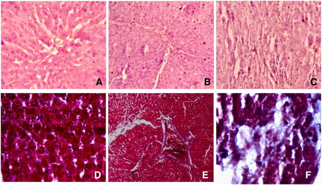
Our results based on the biochemical parameters and HSI are consistent with previous reports in which NDEA or NDMA shows a significant increase in LFT proteins and altered HSI due to hepatic intoxication (Kumar and Kuttan, 2000 , George et al., 2001 , George et al., 2004 , Rezai et al., 2013 , Ahmad et al., 2009 , Ahmad et al., 2011 , Ahmad et al., 2014 , Ahmad and Ahmad, 2012 , Ahmad and Ahmad, 2014 and Latha and Latha, 2014 ). These data clearly demonstrate that NDEA damages the lobular architecture in a dose-dependent manner. The photomicrograph record of control liver sections shows a normal lobular architecture with central veins and radiating hepatic cords (Fig. 1 A and D). Noticeable haemorrhage, dilated central veins, and lymphocyte infiltration were the features of day-7 NDEA treated specimens, whereas day-14 treated animal specimens showed intense neutrophil infiltration, severe haemorrhage and amassing of collagen (Fig. 1 B–C and E–F). The chronic toxicity due to NDEA is attributed to its conversion into an active ethyl radical metabolite by cytochrome P450 (CYP 2E1), which reacts with DNA to cause mutations and carcinogenesis (Anis et al., 2001 ). Therefore, it is proposed that NDEA, similar to other nitroso compounds, such as NDMA, disturbs the redox potential of the cell and may use a similar molecular mechanism to induce hepatic damage in rats.
|
|
|
Fig. 1. Photomicrographs of liver specimens stained with H&E (A–C) and Masson' strichrome (D–F) during NDEA-induced liver injury in rats. (A & D) Control liver with a normal lobular architecture. (B & E) Day-7 NDEA-treated liver specimens. (C & F) Day-14 NDEA-treated liver specimens. |
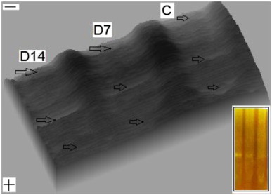
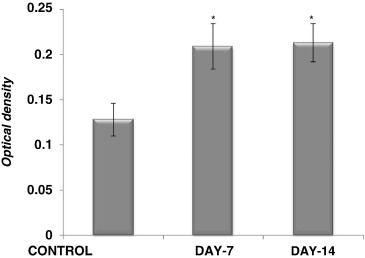
The results of this study further show that the levels of G6PD significantly vary between the control and NDEA treated groups. Upon increasing the contrast of the gels, the software analysis demonstrates the highest levels of G6PD in the day-14 NDEA-treated group, which received a dose of 100 mg kg− 1 b.wt. week− 1 for two weeks (Fig. 2 ). Our spectrophotometry data also support the above findings based on the software analysis of G6PD zymograms and show an approximately 1.63 × and 1.66 × fold increase in the day-7 and day-14 NDEA-treated groups, respectively. A few studies reported interesting findings in which there is apparent controversy for the levels of G6PD under conditions of oxidative stress (Sanz et al., 1997 , Abd Ellah et al., 2004 , Banu et al., 2009 and Taniguchi et al., 1999 ). It has been shown that in AFB1 administered rats (state of oxidative stress), the levels of glutathione metabolizing enzymes, such as G6PD, decrease as a result of the deficient flow of G-6-P through the hexose monophosphate shunt and decreased supply of reduced NADPH for the conversion of GSSG to GSH, thereby causing a switch in the NADP+ /NADPH ratio in favour of NADP+ (Banu et al., 2009 ).
|
|
|
Fig. 2. 3-D surface view of the G6PD zymograms analysed using the Scion Imaging software program. Inset shows zymograms of the corresponding lanes of hepatic G6PD developed by substrate staining. (Symbols − & + shows the direction of the electrophoretic run and the arrow indicates the activity zone of G6PD). |
The literature suggests that under proliferative conditions, G6PD gene expression increases both in foetal hepatocytes (Molero et al., 1994 ) and in cultured mature liver cells (Stanton et al., 1991 ). However, the glucose level in rat livers was decreased during the development of liver cirrhosis if routed for fatty acid and pyruvate synthesis. The glucose channelled to pentose phosphates and reducing equivalents supports DNA synthesis, DNA repair and detoxification reactions (Sanz et al., 1997 ). Our results indicate a significant quantitative differential increase (Fig. 3 ) in the activity of G6PD during NDEA treatment, which may be a consequence of one or both roles of the enzyme: (1) G6PD is required to provide NADPH for microsomal detoxifying mechanisms and/or (2) G6PD provides ribose-5-phosphate for DNA synthesis and repair. Further studies are warranted to evaluate the molecular mechanism of the changes in the G6PD levels under oxidative assault and xenobiotics biotransformation in the damaged liver.
|
|
|
Fig. 3. The bar diagram illustrates the spectrophotometry-based quantitative differences in the levels of G6PD between control and treated groups during NDEA-induced hepatic damage (*P < 0.05). |
Conflict of Interest
None.
Acknowledgements
Financial assistance from Council of Science & Technology [[[#gts0005|CST/SERPD/D-368]] ], UP to RA is gratefully acknowledged for performing this work. The authors sincerely thank the Chairman, Department of Zoology for the necessary facilities.
References
- Abd Ellah et al., 2004 M.R. Abd Ellah, K. Nishimori, M. Goryo, K. Okada, J. Yasuda; Glutathione peroxidase and glucose-6-phosphatedehydrogenase activities in bovine blood and liver; J. Vet. Med. Sci., 66 (2004), pp. 1219–1221
- Ahmad and Ahmad, 2012 A. Ahmad, R. Ahmad; Understanding the mechanism of hepatic fibrosis and potential therapeutic approaches; Saudi J. Gastroenterol., 18 (2012), pp. 155–167
- Ahmad and Ahmad, 2014 A. Ahmad, R. Ahmad; Resveratrol mitigate structural changes and hepatic stellate cell activation in N′-Nitrosodimethylamine-induced liver fibrosis via restraining oxidative damage ; Chem. Biol. Interact., 221 (2014), pp. 1–12
- Ahmad and Hasnain, 2005 R. Ahmad, A. Hasnain; Ontogenetic changes and developmental adjustments in lactate dehydrogenase isozymes of an obligate air-breathing fish Channa punctatus during deprivation of air access. Comp. Biochem. Physiol., Part B: biochem ; Mol. Biol., 140 (2005), pp. 271–278
- Ahmad and Hasnain, 2006 R. Ahmad, A. Hasnain; Differential expressions of G6PD and alkaline phosphatase isozymes associated with ontogeny and air-breathing transition in Channa punctata; Asian Fish. Sci., 19 (2006), pp. 141–148
- Ahmad et al., 2009 R. Ahmad, S. Ahmed, N.U. Khan, A. Hasnain; Operculina turpethum attenuates Nʹ-nitrosodimethylamine induced toxic liver injury and clastogenicity in rats ; Chem. Biol. Interact., 181 (2009), pp. 145–153
- Ahmad et al., 2011 A. Ahmad, R. Fatima, V. Maheshwari, R. Ahmad; Effect of Nʹ-nitrosodimethylamine on red blood cell rheology and proteomic profiles of brain in male albino rats; Interdiscip. Toxicol., 4 (2011), pp. 125–131
- Ahmad et al., 2014 A. Ahmad, N. Afroz, U.D. Gupta, R. Ahmad; Vitamin B12 supplement alleviates Nʹ-nitrosodimethylamine-induced hepatic fibrosis in rats; Pharm. Biol., 52 (2014), pp. 516–552
- Anis et al., 2001 K.V. Anis, N.V.R. Kumar, R. Kuttan; Inhibition of chemical carcinogenesis by biberine in rats and mice; J. Pharm. Pharmacol., 53 (2001), pp. 763–768
- Banu et al., 2009 G.S. Banu, G. Kumar, A.G. Murugesan; Ethanolic leaves extract of Trianthema portulacastrum L . ameliorates aflatoxin B1 induced hepatic damage in rats ; Indian J. Clin. Biochem., 24 (2009), pp. 250–256
- Biagiotti et al., 2000 E. Biagiotti, K.S. Bosch, P. Ninfali, W.M. Frederiks, C.J.F.V. Noorden; Post-translational regulation of glucose-6-phosphate dehydrogenase activity in tongue epithelium; J. Histochem. Cytochem., 48 (2000), pp. 971–977
- Farazi and De Pinho, 2006 P.A. Farazi, R.A. De Pinho; Hepatocellular carcinoma pathogenesis: from genes to environment; Nat. Rev. Cancer, 6 (2006), pp. 674–687
- Fitch et al., 1959 W.M. Fitch, R. Hill, I.L. Chaikoff; Hepatic glycolytic enzyme activities in the Alloxan-diabetic rat: Response to glucose and fructose feeding; J. Biol. Chem., 234 (1959), pp. 2811–2813
- Frederiks et al., 2003 W.M. Frederiks, K.S. Bosch, J.S.S.G. De Jong, C.J.F.V. Noorden; Post- translational regulation of glucose-6-phosphate dehydrogenase activity in (pre) neoplastic lesions in rat liver; J. Histochem. Cytochem., 51 (1) (2003), pp. 105–112
- George et al., 2001 J. George, J.R. Rao, S. Stern, G. Chandrakasan; Dimethylnitrosamine-induced liver injury in rats: the early deposition of collagen; Toxicology, 156 (2001), pp. 129–138
- George et al., 2004 J. George, M. Tsutsumi, S. Takase; Expression of hyaluronic acid in N-nitrosodimethylamine induced hepatic fibrosis in rats; Int. J. Biochem. Cell Biol., 36 (2) (2004), pp. 307–319
- Glock and Mc Lean, 1955 G.E. Glock, P. McLean; A preliminary investigation of the hormonal control of the hexose monophosphate oxidative pathway; Biochem. J., 61 (1955), pp. 390–397
- Holten et al., 1976 D. Holten, D. Procsal, H.L. Chang; Regulation of pentose phosphate pathway dehydrogenases by NADP+ /NADPH ratios ; Biochem. Biophys. Res. Commun., 68 (1976), pp. 436–441
- Ibrahim et al., 2014 M.A. Ibrahim, A.H.M. Ghazy, M.H.S. Ahmed, M.A. Ghazy, M.M.A. Monsef; Purification and characterization of glucose-6-phosphate dehydrogenase from camel liver; Enzym. Res. (2014), pp. 1–10
- Jain et al., 2003 M. Jain, D.A. Brenner, L. Cui, C.C. Lim, B. Wang, D.R. Pimentel, K. Stanley, D.B. Sawyer, J.A. Leopold, D.E. Handy, J. Loscalzo, C.S. Apstein, R. Liao; Glucose-6-phosphate dehydrogenase modulates cytosolic redox status and contractile phenotype in adult cardiomyocytes; Circ. Res., 93 (2) (2003), pp. 1–11
- Katsurada et al., 1989 A. Katsurada, N. Iritani, H. Fukuda, Y. Matsumura, T. Noguchi, T. Tanaka; Effects of nutrients and insulin on transcriptional and post-transcriptional regulation of glucose-6-phosphate dehydrogenase synthesis in rat liver; Biochim. Biophys. Acta, 1006 (1989), pp. 104–110
- Kletzein et al., 1994 R.F. Kletzein, P.K.W. Harris, L.A. Foellmi; Glucose-6-phopsphate dehydrogenase: a ʹhousekeepingʹ enzyme subject to tissue-specific regulation by hormones, nutrients, and oxidant stress; FASEB J., 8 (1994), pp. 174–181
- Köhler and Noorden, 2003 A. Köhler, C.J.F.V. Noorden; Reduced nicotinamide adenine dinucleotide phosphate and the higher incidence of pollution-induced liver cancer in female flounder; Environ. Toxicol. Chem., 22 (2003), pp. 2703–2710
- Kumar and Kuttan, 2000 R.N.V. Kumar, R. Kuttan; Inhibition of Nʹ-nitrosodiethylamine-induced hepatocarcinogenesis by Picroliv; J. Exp. Clin. Cancer Res., 19 (4) (2000), pp. 459–465
- Latha and Latha, 2014 B. Latha, M.S. Latha; Preventive effect of Leucas aspera methanolic extract on Nʹ-Nitrosodiethylamine induced sub-acute liver toxicity in male wistar rats ; Int. J. Pharm. Sci. Res., 5 (6) (2014), pp. 2349–2353
- Linza et al., 2013 A. Linza, P.J. Wills, P.N. Ansil, S.P. Prabha, A. Nitha, B. Latha, K.O. Sheeba, M.S. Latha; Dose–response effects of Elephantopus scaber methanolic extract on N-nitrosodiethylamine-induced hepatotoxicity in rats ; Chin. J. Nat. Med., 11 (4) (2013), pp. 0362–0370
- Lowry et al., 1951 O.H. Lowry, N.J. Rosebrough, A.L. Farr, R.J. Randall; Protein measurement with Folin phenol reagent; J. Biol. Chem., 193 (1951), pp. 265–275
- Molero et al., 1994 C. Molero, M. Benito, M. Lorenzo; Glucose-6-phosphate dehydrogenase gene expression in fetal hepatocyte primary cultures under non-proliferative and proliferative conditions; Exp. Cell Res., 210 (1994), pp. 26–32
- Nitha et al., 2014 A. Nitha, S.P. Prabha, P.N. Ansil, M.S. Latha; Curative Effect of Woodfordia fruticosa kurz flowers on N-nitrosodiethylamine induced hepatocellular carcinoma in rats ; Int. J. Pharm. Sci., 6 (2) (2014), pp. 150–155
- Pandolfi et al., 1995 P.P. Pandolfi, F. Sonati, R. Rivil, P. Mason, F. Grosveld, L. Luzzattol; Targeted disruption of the housekeeping gene encoding glucose 6-phosphate dehydrogenase (G6PD): G6PD is dispensable for pentose synthesis but essential for defense against oxidative stress; EMBO J., 14 (21) (1995), pp. 5209–5215
- Rezai et al., 2013 A. Rezai, A. Fazlara, M. Haghikaramolah, A. Shahriari, H.N. Zadeh, M. Pashforosh; Effect of Echinacea purpurea on hepatic and renal toxicity induced by diethylnitrosamine in rats ; Jund. J. Nat. Pharm. Prod., 8 (2) (2013), pp. 60–64
- Rudack et al., 1971a D. Rudack, E.M. Chisholm, D. Holten; Rat liver glucose 6-phosphate dehydrogenase. Regulation by carbohydrate diet and insulin; J. Biol. Chem., 246 (1971), pp. 1249–1254
- Rudack et al., 1971b D. Rudack, E.M. Gozukara, E.M. Chisholm, D. Holten; The effect of dietary carbohydrate and fat on the synthesis of rat liver 6-phosphogluconate dehydrogenase; Biochim. Biophys. Acta, 252 (2) (1971), pp. 305–313
- Sanz et al., 1997 N. Sanz, C. Díez-Fernández, A.M. Valverde, M. Lorenzo, M. Benito, M. Cascales; Malic enzyme and glucose-6-phosphate dehydrogenase gene expression increases in rat liver cirrhogenesis; Br. J. Cancer, 75 (1997), pp. 487–492
- Shannon et al., 2000 A.F.W. Shannon, S. Gover, V.M. Lam, M.J. Adams; Human glucose-6-phosphate dehydrogenase: the crystal structure reveals a structural NADP+ molecule and provides insights into enzyme deficiency ; Structure, 8 (2000), pp. 293–303
- Shaw and Prasad, 1970 C.R. Shaw, R. Prasad; Starch gel electrophoresis of enzymes: a compilation of recipes; Biochem. Genet., 4 (1970), pp. 279–320
- Stanton, 2012 R.C. Stanton; Glucose-6-phosphate dehydrogenase, NADPH, and cell survival; IUBMB Life, 64 (5) (2012), pp. 362–369
- Stanton et al., 1991 R.C. Stanton, J.L. Sciffer, D.C. Boxer, E. Zimmermann, L.C. Cantlet; Rapid release of bound glucose-6-phosphate dehydrogenase by growth factors; J. Biol. Chem., 266 (1991), pp. 12442–12448
- Taniguchi et al., 1999 M. Taniguchi, A. Yasutake, K. Takedomi, K. Inoue; Effects of N-nitrosodimethylamine (NDMA) on the oxidative status of rat liver; Arch. Toxicol., 73 (1999), pp. 141–146
- Tepperman and Tepperman, 1958 H.M. Tepperman, J. Tepperman; The hexose monophosphate shunt and adaptive hyperlipogenesis; Diabetes, 7 (6) (1958), pp. 478–485
- Tian et al., 1998 W.N. Tian, L.D. Braunstein, J. Pang, K.M. Stuhlmeier, Q.X. Chao, X. Tian, R.C. Stanton; Importance of glucose-6-phosphate dehydrogenase activity for cell growth; J. Biol. Chem., 273 (17) (1998), pp. 10609–10617
- Ulusu and Tandogan, 2006 N.N. Ulusu, B. Tandogan; Purification and kinetics of sheep kidney cortex glucose-6-phosphate dehydrogenase; Comp. Biochem. Physiol. B: Biochem. Mol. Biol., 143 (2) (2006), pp. 249–255
- Wanatabe and Taketa, 1972 A. Wanatabe, K. Taketa; Purification of rat liver glucose-6-phosphate dehydrogenase by batch wise adsorption and substrate elution; J. Biochem., 72 (1972), p. 1277
- Watanabe and Taketa, 1973 A. Watanabe, K. Taketa; Immunological studies on glucose-6-phosphate dehydrogenase of rat liver; Arch. Biochem. Biophys., 158 (1973), pp. 43–52
- Wills et al., 2006 P.J. Wills, V. Suresh, M. Arun, V.V. Asha; Antiangiogenic effect of Lygodium flexuosum against N-nitrosodiethylamine-induced hepatotoxicity in rats ; Chem. Biol. Interact., 164 (1–2) (2006), pp. 25–38
- Zhang et al., 2012 C.L. Zhang, T. Zheng, X.L. Zhao, L.H. Yu, Z.P. Zhu, K.Q. Xie; Protective effects of garlic oil on hepatocarcinoma induced by Nʹ-nitrosodietylamine in rats; Int. J. Biol. Sci. (2012), pp. 363–374
Document information
Published on 27/03/17
Licence: Other
Share this document
Keywords
claim authorship
Are you one of the authors of this document?


